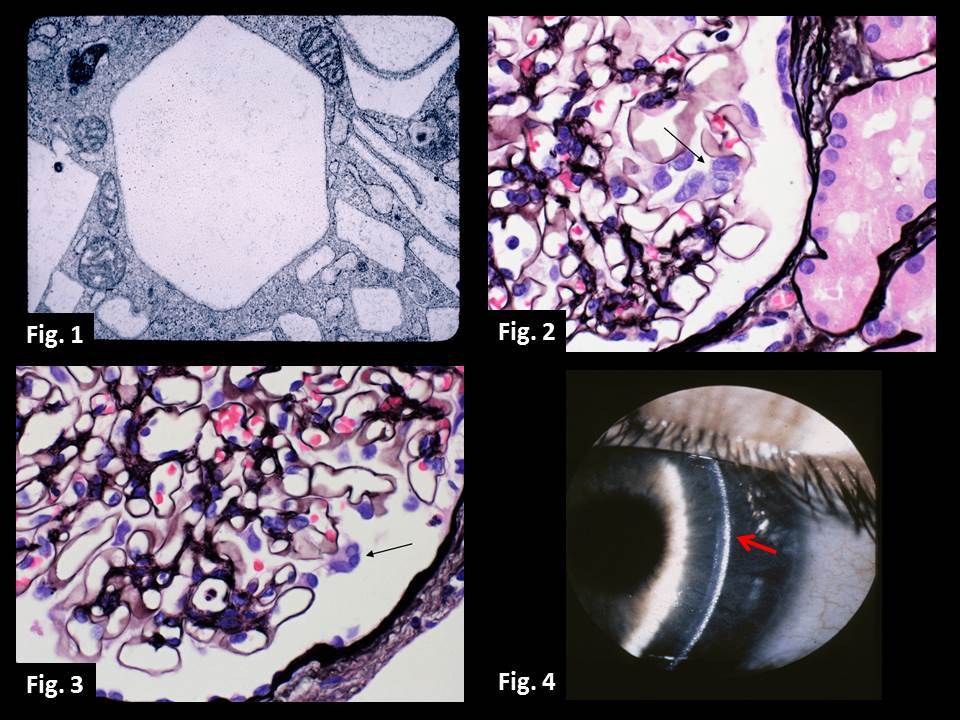
Cystinosis is an autosomal recessive lysosomal storage disease caused by CTNS gene mutations. The normal product of the CTNS gene is cystinosin, a transport protein that moves the amino acid cystine out of the lysosome. CTNS gene mutations (more than 80 have been described thus far) result in the accumulation of cystine within lysosomes. Cystine crystals, which accumulate in various tissues such as the kidney and eye, can be detected using polarization or electron microscopy (Fig. 1). In the kidney, multinucleated podocytes (Fig. 2-3) and increased numbers of atubular glomeruli (not shown) may be identified by routine light microscopy. In the eye, corneal opacities may be seen using slit lamp examination (Fig. 4). Most cases of cystinosis tend to be infantile; however, juvenile and adult forms of the disease are also recognized. The diagnosis is confirmed using genetic testing.
References
Larsen CP, Walker PD, Thoene JG. The incidence of atubular glomeruli in nephropathic cystinosis renal biopsies. Mol Genet Metab. 2010 Dec;101(4):417-20. PMID: 20826102.
David G. Cogan Ophthalmic Pathology Collection, NEI/NIH (Fig. 1 and 4).
Quick note: This post is to be used for informational purposes only and does not constitute medical or health advice. Each person should consult their own doctor with respect to matters referenced. Arkana Laboratories assumes no liability for actions taken in reliance upon the information contained herein.


