
Did you know there could be other uses for Congo Red Stain?
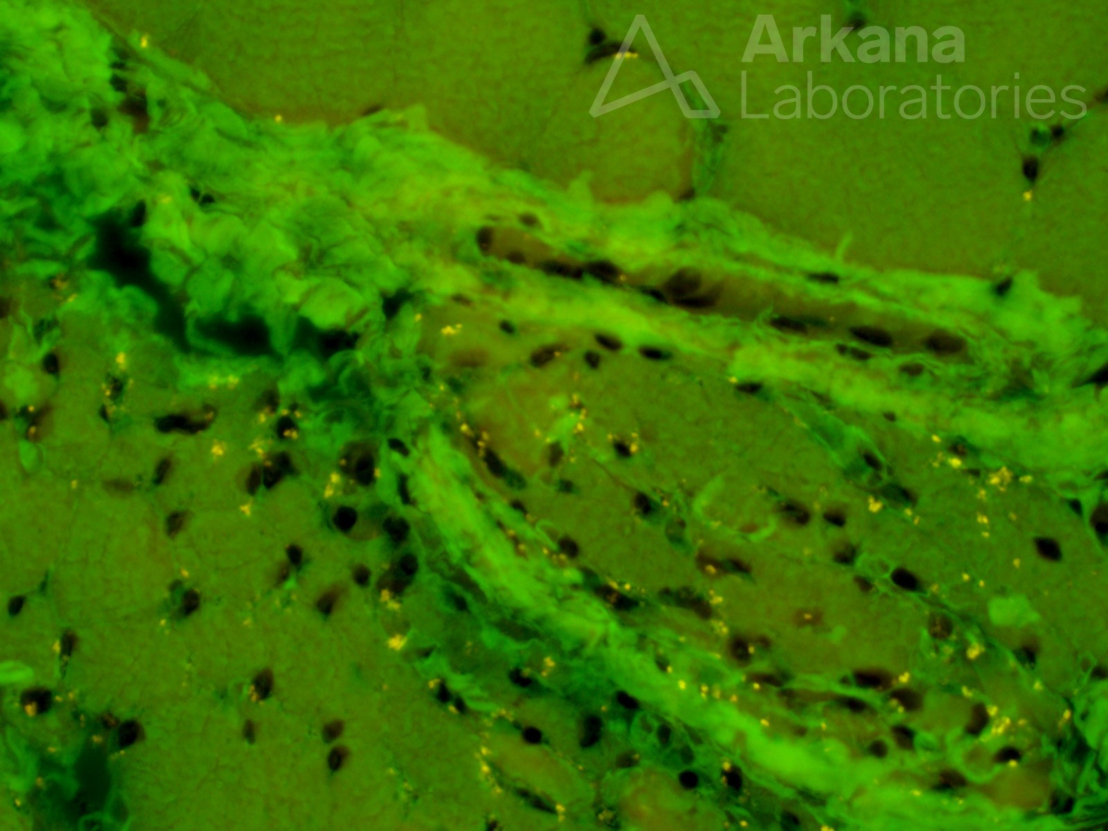
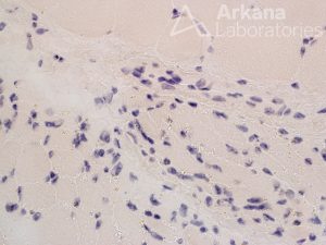
Additional unintended useful information can be obtained from many histochemical stains used in the routine evaluation of skeletal muscle biopsies. In addition to identifying amyloid deposition, Congo red stain provides some morphologic detail. In this case, granular brown lipofuscin pigment can be seen in the atrophic muscle fibers under routine light microscopy. The lipofuscin pigment is also autofluorescent, showing a white-yellow color under fluorescence microscopy.
Congo Red stained frozen section of skeletal muscle under routine light microscopy (top) and fluorescence microscopy with FITC filter (bottom); original magnification: 400x each.
Quick note: This post is to be used for informational purposes only and does not constitute medical or health advice. Each person should consult their own doctor with respect to matters referenced. Arkana Laboratories assumes no liability for actions taken in reliance upon the information contained herein.

