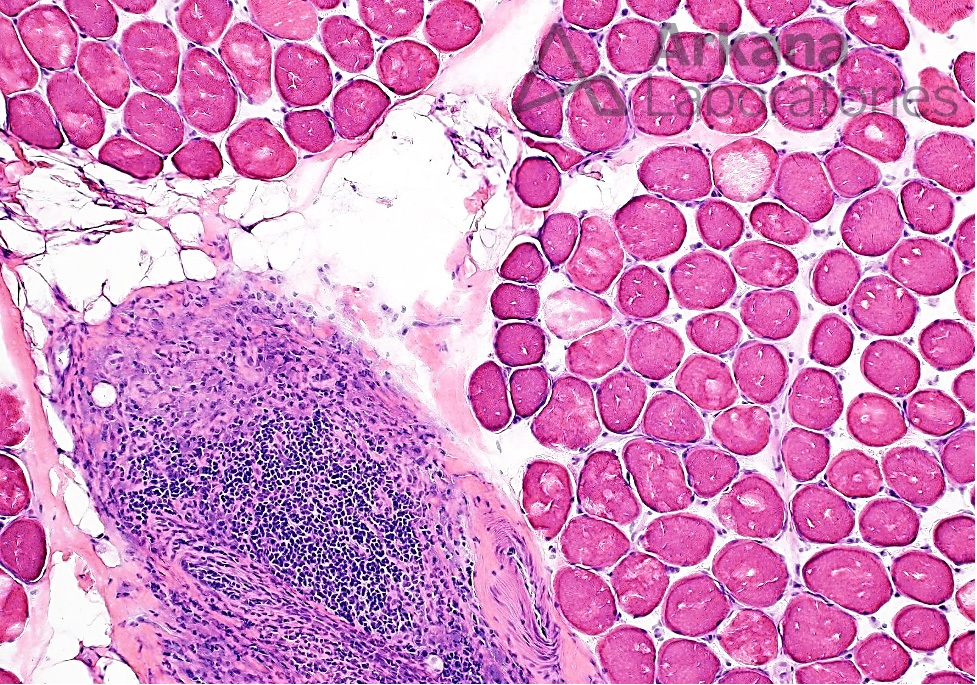
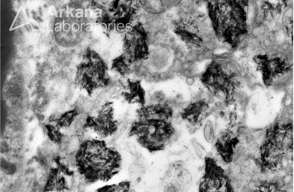
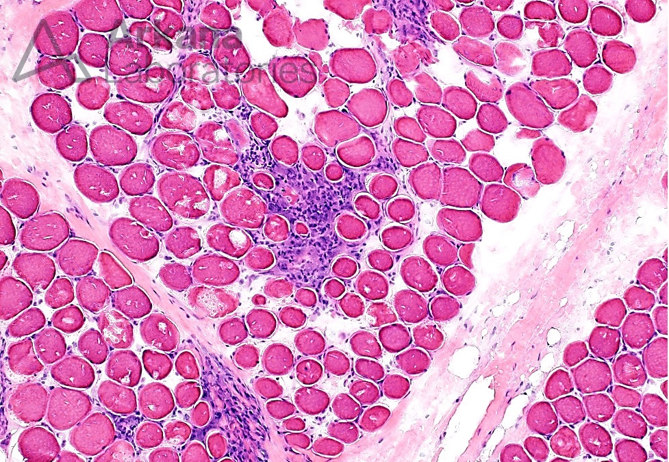
This previously healthy pediatric patient presented with peculiar facial muscle hypertrophy, and genetic testing revealed one pathologic variant and two variants of uncertain significance (VUSs). A muscle biopsy of the thigh was performed to further evaluate this patient. Frozen sections and ultrastructural examination images of the inflammatory infiltrate are shown below. What is your diagnosis?
Answer: Macrophagic Myofasciitis (MMF)
The muscle biopsy demonstrates a few endomysial and perimysial foci of mild chronic inflammation and associated variably frequent collections of CD68-positive histiocytic cells with moderate to abundant amounts of pale basophilic cytoplasm. No significant associated muscle fiber injury is noted. The chronic lymphocytic inflammatory cells are predominantly comprised of CD8-positive T-cells. The overall morphologic features are consistent with Macrophagic Myofasciitis (MMF), a relatively uncommon localized inflammatory process related to prior alum (aluminum) containing vaccines. See references.
References:
Chkheidze R, et al. Morin stain detects aluminum-containing macrophages in macrophagic myofasciitis and vaccination granuloma with high sensitivity and specificity. J Neuropathol Exp Neurol. 2017;76:323-331.
Gherardi RK, Authier FJ. Macrophagic myofasciitis: characterization and pathophysiology. Lupus. 2012 Feb;21(2):184-189.
Israeli E, Agmon-Levin N, Blank M, Shoenfeld Y. Macrophagic myofaciitis a vaccine (alum) autoimmune-related disease. Clin Rev Allergy Immunol. 2011;41(2):163-168.
Quick note: This post is to be used for informational purposes only and does not constitute medical or health advice. Each person should consult their own doctor with respect to matters referenced. Arkana Laboratories assumes no liability for actions taken in reliance upon the information contained herein.


