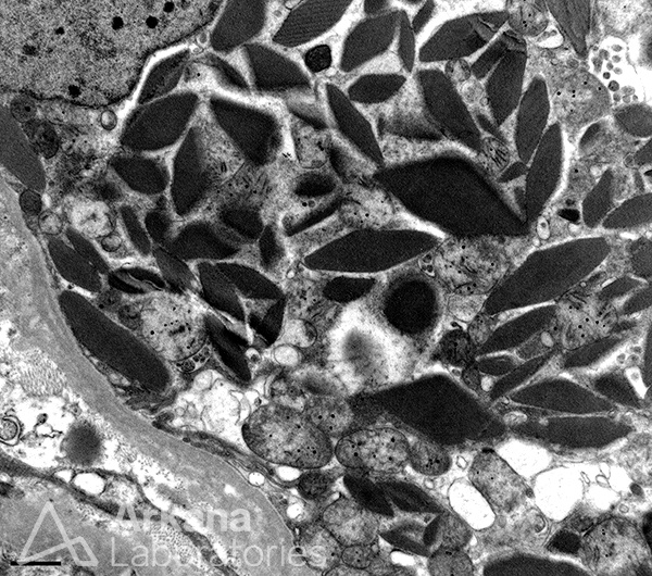
What is the most likely diagnosis in a patient with an abnormal serum protein electrophoresis?
Answer: The image depicts numerous diamond to rhomboid-shaped crystals within the cytoplasm of a proximal tubular epithelial cell and in the correct context of light chain restriction by immunofluorescence, is consistent with a diagnosis of light chain proximal tubulopathy with crystals. By light microscopy, numerous proximal tubules demonstrated intracytoplasmic crystals which were characteristically fuchsinophilic on the Masson’s trichrome stain. By immunofluorescence, the proximal tubules demonstrated kappa light chain restriction and clinically the patient was found to have multiple myeloma on a follow-up bone marrow biopsy. Of note, some cases will not show expected light chain restriction on routine immunofluorescence. In these cases, paraffin immunofluorescence with protease digestion is a useful tool to help reveal the light chain restriction.
Quick note: This post is to be used for informational purposes only and does not constitute medical or health advice. Each person should consult their own doctor with respect to matters referenced. Arkana Laboratories assumes no liability for actions taken in reliance upon the information contained herein.

