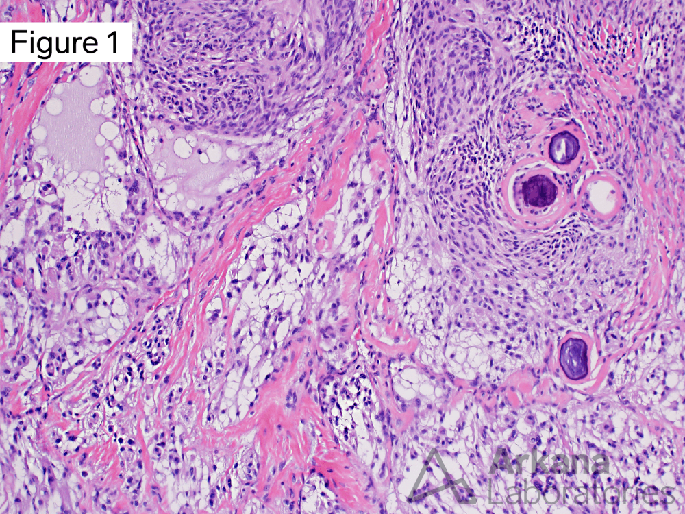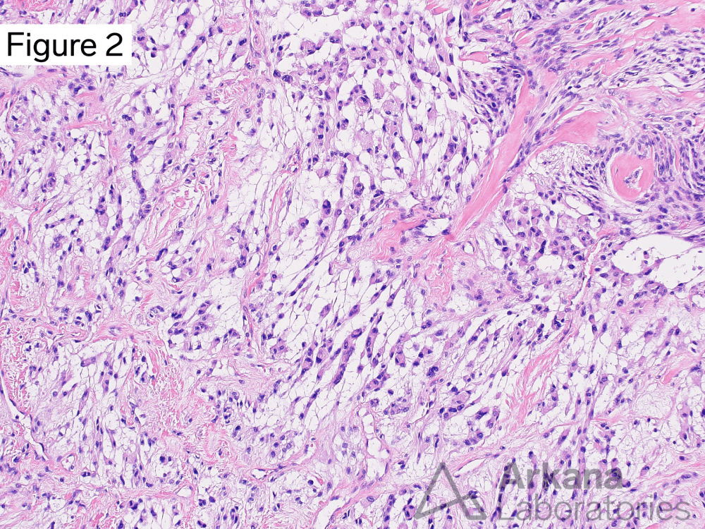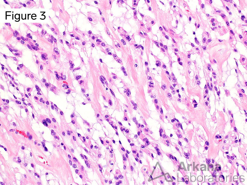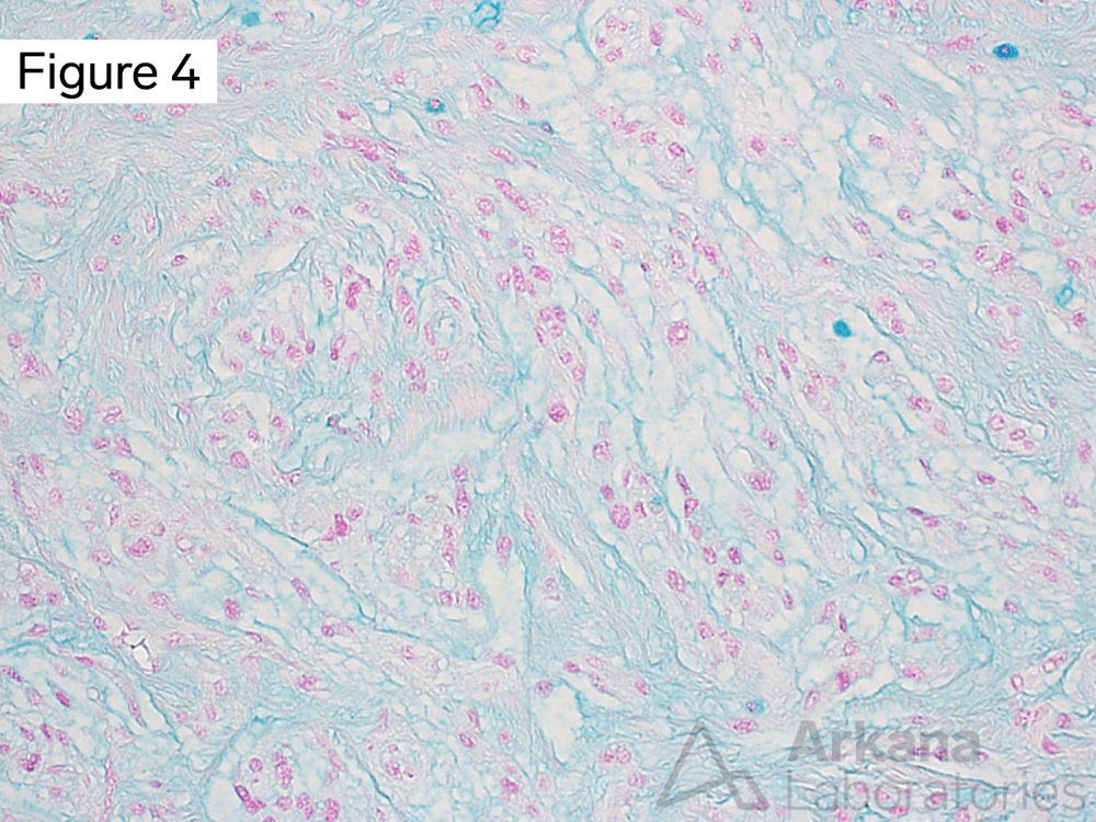
This 50-year-old patient presented with visual changes and word-finding difficulties. Brain MRI demonstrated a 4.6 x 4.5 x 3.5 cm well-circumscribed contrast-enhancing dural-based extra-axial mass, with dural tail and mass effect on the left frontal lobe. The lesion was gross totally resected.
What is your diagnosis based on Figures #1 through #4?
A. Chordoid meningioma
B. Pilocytic astrocytoma
C. Glioblastoma
D. Lymphoma
Answer: Chordoid meningioma
The morphologic features are consistent with the pathologic diagnosis of chordoid meningioma. This meningioma subtype has a propensity for local recurrence and is therefore considered a WHO grade 2 lesion.
The overall morphologic features of this dural-based extra-axial mass are not those of glioblastoma, pilocytic astrocytoma, or lymphoma. Other considerations, in this case, include tumor-to-meningioma metastasis (a rare phenomenon), chordoma (cytokeratin, S100 protein, and brachury positive), and chordoid glioma of the third ventricle (GFAP and TTF1 positive).
References/Additional Reading
Sangoi AR, Dulai MS, Beck AH, Brat DJ, Vogel H. Distinguishing chordoid meningiomas from their histologic mimics: an immunohistochemical evaluation. Am J Surg Pathol. 2009 May;33(5):669-81. doi: 10.1097/PAS.0b013e318194c566. PMID: 19194275; PMCID: PMC4847145.
Couce ME, Aker FV, Scheithauer BW. Chordoid meningioma: a clinicopathologic study of 42 cases. Am J Surg Pathol. 2000 Jul;24(7):899-905. doi: 10.1097/00000478-200007000-00001. Erratum in: Am J Surg Pathol 2000 Sep;24(9):1316-7. PMID: 10895812.
Quick note: This post is to be used for informational purposes only and does not constitute medical or health advice. Each person should consult their own doctor with respect to matters referenced. Arkana Laboratories assumes no liability for actions taken in reliance upon the information contained herein.


