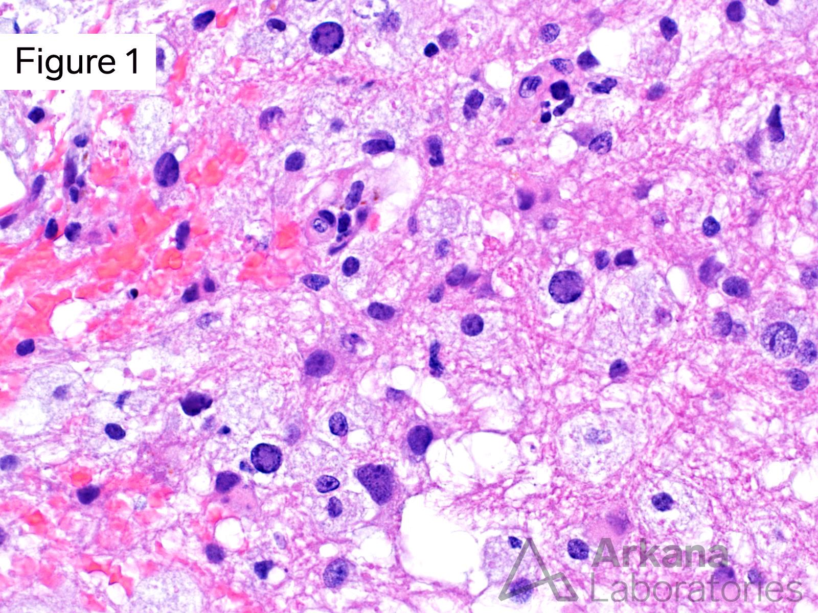
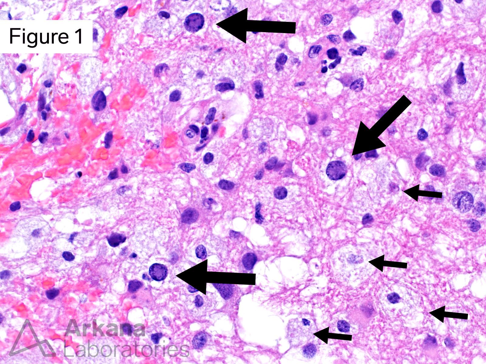
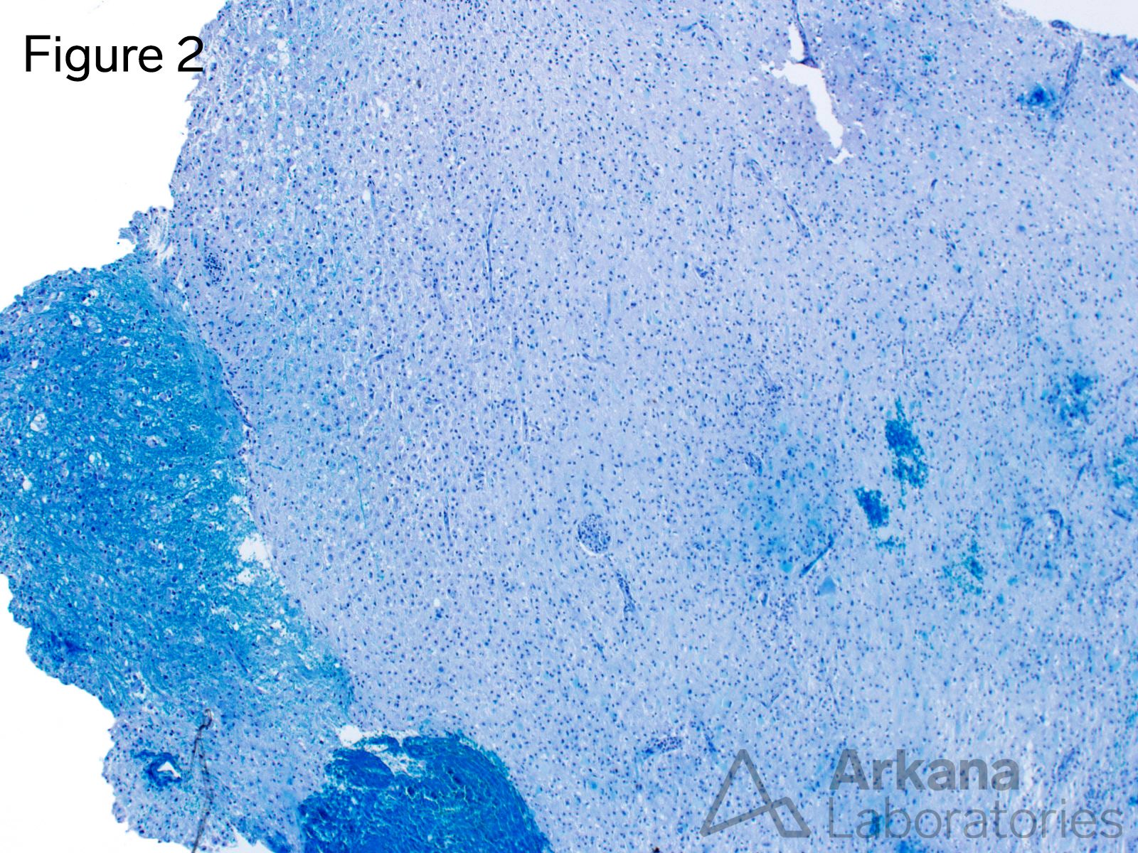
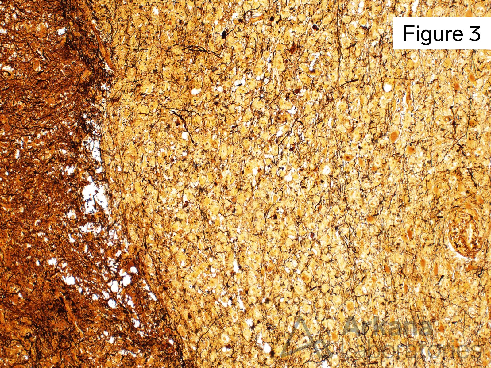
This 60-year-old immunocompromised patient presented with a one-week history of progressive right-sided weakness, difficulty concentrating, and impaired word finding ability. Brain MRI showed a 5.5 x 4.0 x 2.7 cm, T2 and FLAIR hyperintense, T1 hypointense, non-contrast enhancing mass involving the white matter of the right frontal lobe.
Based on Figures #1 – #4, what stain would you like to perform to further evaluate this patient’s mass lesion?
A. Polyomavirus IHC
B. IDH1 R132H IHC
C. Cytokeratin IHC
D. AFB stain
Answer: Polyomavirus IHC
Given the presence of morphologic changes of a demyelinating lesion and oligodendrocytes with intranuclear viral cytopathic change, performing a polyomavirus immunohistochemical stain to detect JC virus is the correct choice.
JC virus is the causative agent for Progressive Multifocal Leukoencephalopathy (PML). Please see additional Reference(s) / additional reading.
The nuclear atypia and elevated proliferative fraction, may be mistaken for a neoplastic process.
Reference(s) / additional reading:
Berger JR, Aksamit AJ, Clifford DB, Davis L, Koralnik IJ, Sejvar JJ, Bartt R, Major EO, Nath A. PML diagnostic criteria: consensus statement from the AAN Neuroinfectious Disease Section. Neurology. 2013 Apr 9;80(15):1430-8. doi: 10.1212/WNL.0b013e31828c2fa1. PMID: 23568998; PMCID: PMC3662270.
Atkinson AL, Atwood WJ. Fifty Years of JC Polyomavirus: A Brief Overview and Remaining Questions. Viruses. 2020 Sep 1;12(9):969. doi: 10.3390/v12090969. PMID: 32882975; PMCID: PMC7552028.
Zu Rhein GM. Ultrastructural studies in progressive multifocal leukoencephalopathy, a demyelinating disease of man of probable viral etiology. Int Arch Allergy Appl Immunol. 1969;36:Suppl:463-87. PMID: 4313668.
Quick note: This post is to be used for informational purposes only and does not constitute medical or health advice. Each person should consult their own doctor with respect to matters referenced. Arkana Laboratories assumes no liability for actions taken in reliance upon the information contained herein.


