Clinical History
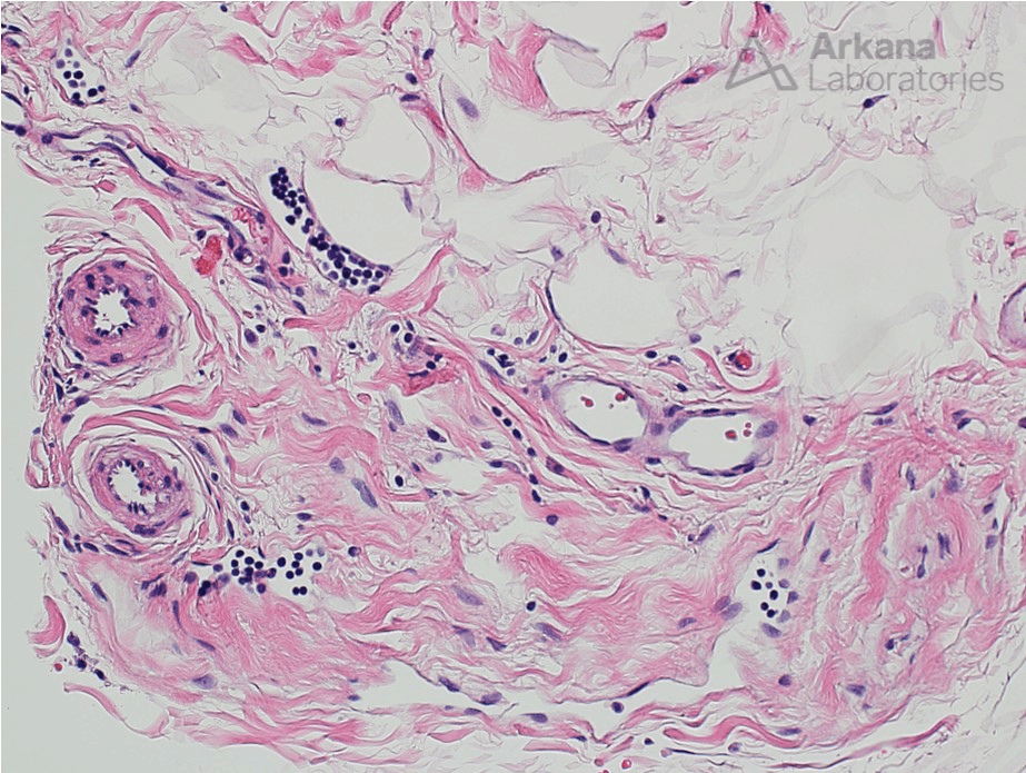
This 50-year-old patient presented with right thigh pain. Their muscle biopsy showed mild denervation type changes. Several small portions of fibroadipose tissue were included in the biopsy.
The small collections of lymphocytes seen in Figure 1 are associated with which of the following?
A. Fasciitis
B. Vasculitis
C. Nerve twigs
D. Lymphatic spaces

Correct Answer: D. Lymphatic spaces
The correct answer is that these lymphocytes are within lymphatic spaces.
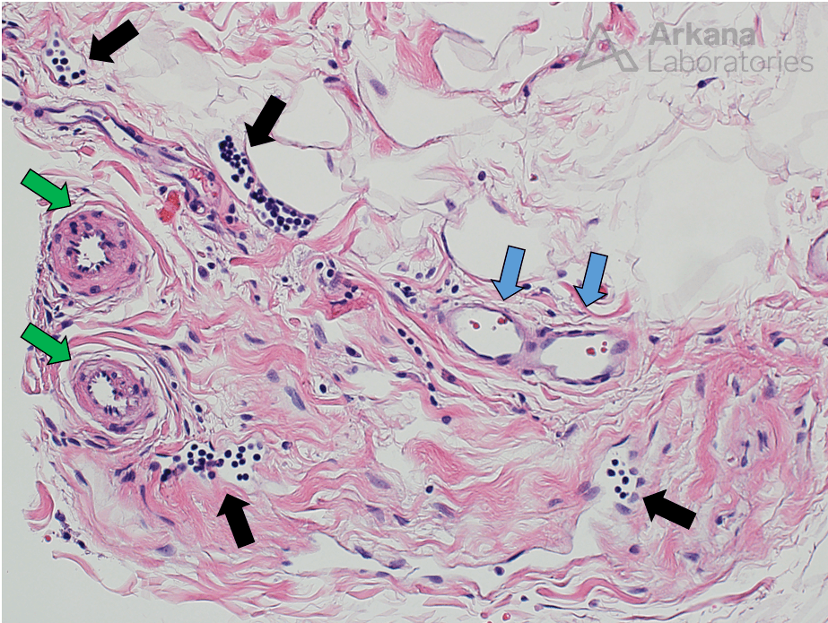
The lymphocytes in this image are present within small irregularly shaped channels lined by endothelial cells with elongate nuclei (lymphatic endothelial cells).
Note the following:
Arterioles green arrows
Venules blue arrows
Lymphatic spaces black arrows

Quick note: This post is to be used for informational purposes only and does not constitute medical or health advice. Each person should consult their own doctor with respect to matters referenced. Arkana Laboratories assumes no liability for actions taken in reliance upon the information contained herein.

