Clinical History
This 80-year old patient presented with new onset seizures. Brain MRI demonstrated a 4.6 x 3.4 x 3.0 cm heterogeneously enhancing left frontal mass with associated mass effect.
The majority of the tumor showed morphologic features characteristic for IDH-wildtype glioblastoma. ATRX nuclear immunostaining was retained, and there was no cytoplasmic staining for IDH1 R132H mutant protein. Many of the neoplastic glial cells showed strong nuclear staining for p53.
What is your diagnosis based on the representative images that were also seen in several foci in this patient’s brain neoplasm?
Answer:
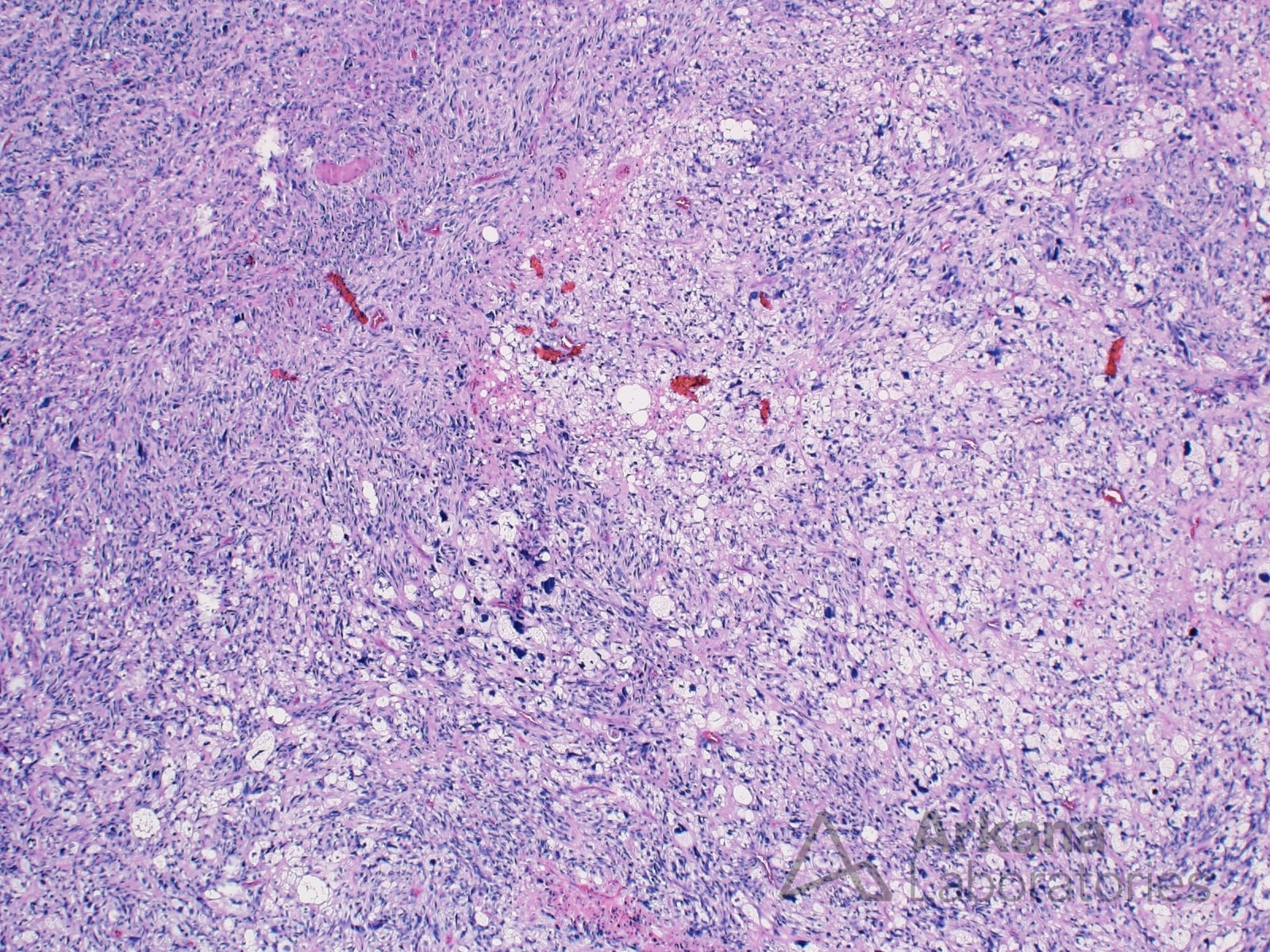
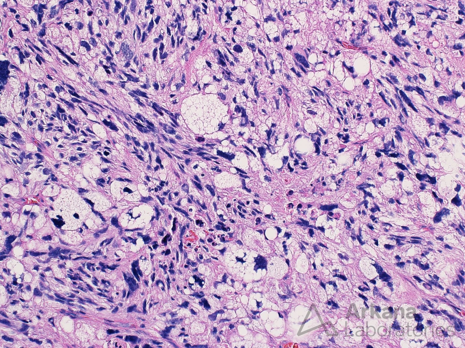
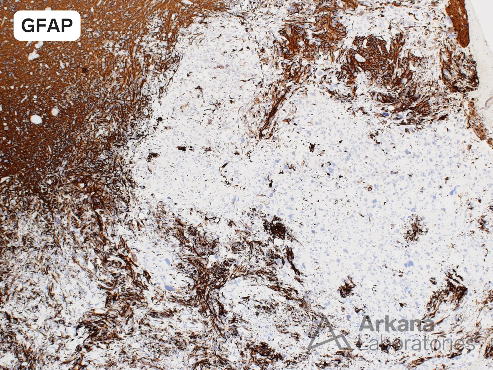
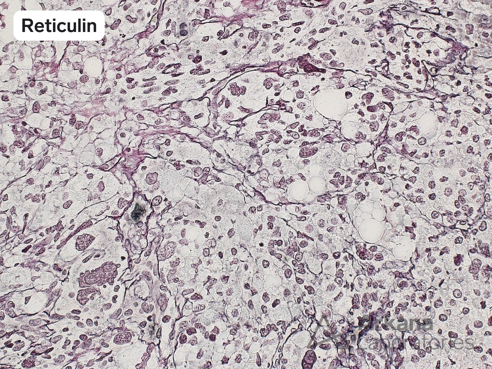
The images demonstrate the focal transition from a high grade glial neoplasm with astrocytic cytomorphology (glioblastoma) to an area showing mesenchymal-type differentiation (liposarcomatous in this case). The cells in this area are negative for GFAP, and there is corresponding deposition of an intercellular reticulin network.
These features are consistent with the presence of an IDH-wildtype glioblastoma with focal areas of mesenchymal metaplasia (liposarcomatous).
The term “gliosarcoma” is reserved for tumors showing prominent/extensive areas of alternating neoplastic glial and mesenchymal differentiation.
References/Additional Readings:
- WHO Classification of Tumours Editorial Board. Central nervous system tumours. (2021), vol 6. WHO Classification of Tumours series., 5 edn. International Agency for Research on Cancer, Lyon (France) pages 47-48
Quick note: This post is to be used for informational purposes only and does not constitute medical or health advice. Each person should consult their own doctor with respect to matters referenced. Arkana Laboratories assumes no liability for actions taken in reliance upon the information contained herein.