Clinical History
This 50-year-old patient was found to have in incidental dural based mass. What is your diagnosis?
Correct Answer
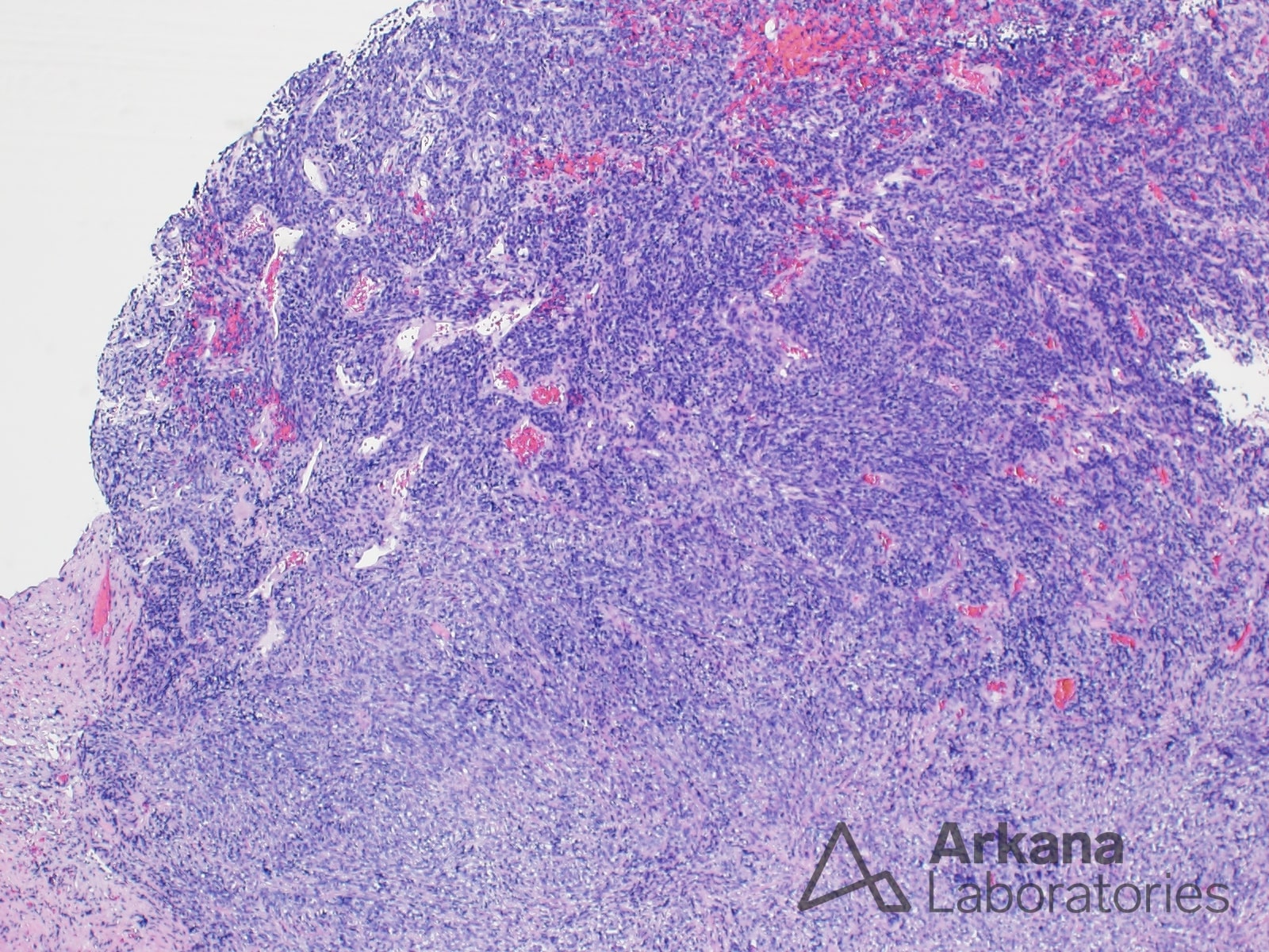
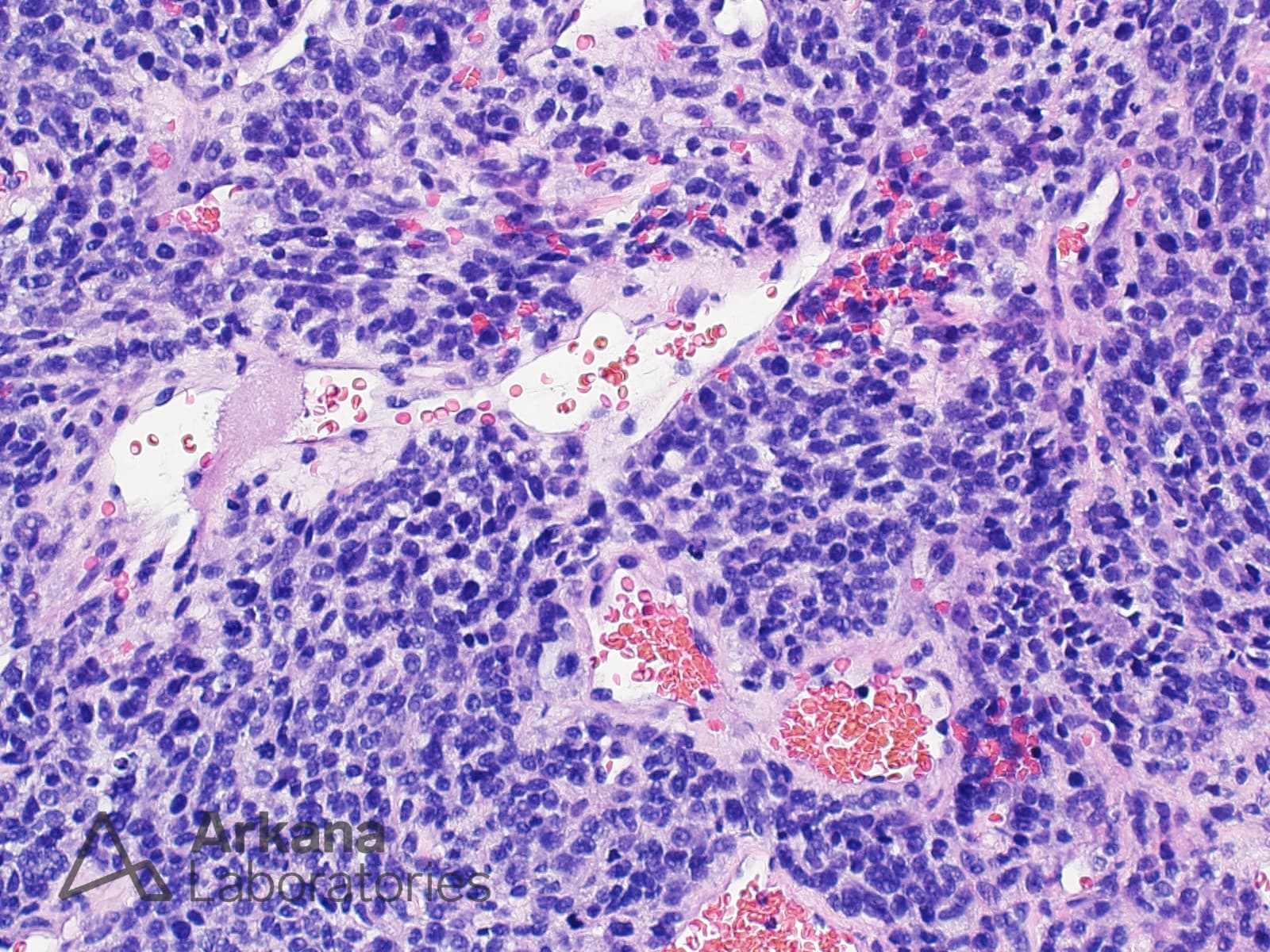
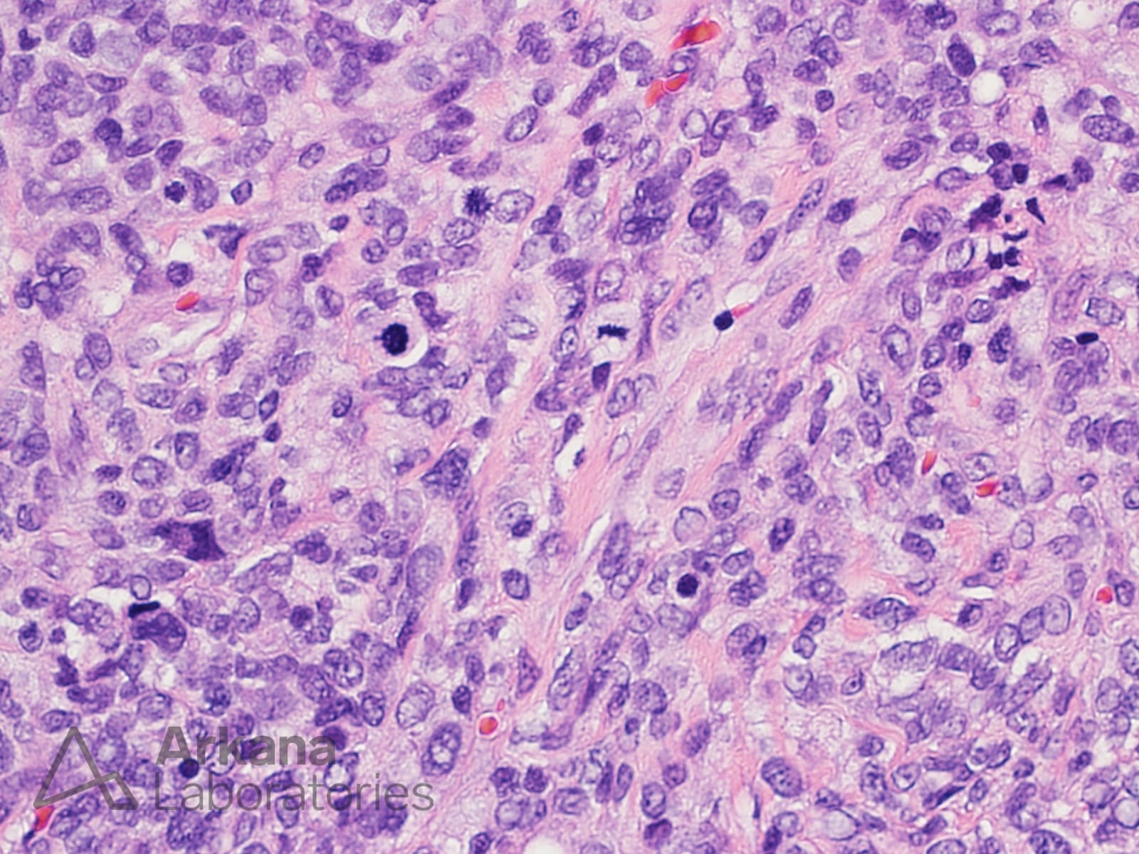
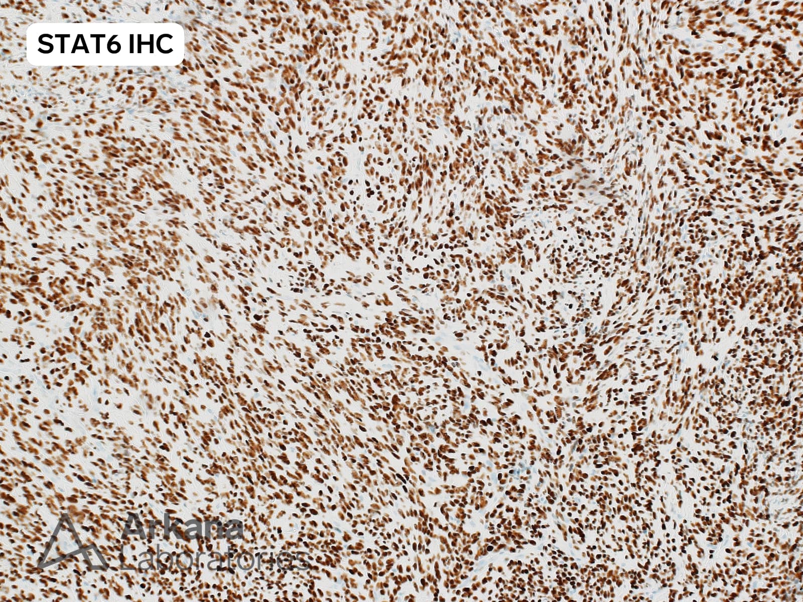
The H&E stained sections demonstrate a cellular proliferation of relatively monotonous appearing spindle to ovoid cells with patchy “staghorn” type vascular pattern. Frequent mitotic figures (>20/10hpf) were present. STAT6 immunostain shows diffuse strong staining of the neoplastic cell nuclei.
The overall features are consistent with the diagnosis of a dural-based solitary fibrous tumor / SFT. This lesion was previously designated as hemangiopericytoma / HPC).
The STAT6 overexpression in the neoplastic cells is driven by the presence of NAB2::STAT6 gene fusion due to genomic inversion at 12q13. This is an excellent example of an immunostain being used to impute a genetic abnormality (i.e. surrogate marker for molecular testing).
Quick note: This post is to be used for informational purposes only and does not constitute medical or health advice. Each person should consult their own doctor with respect to matters referenced. Arkana Laboratories assumes no liability for actions taken in reliance upon the information contained herein.