Clinical History
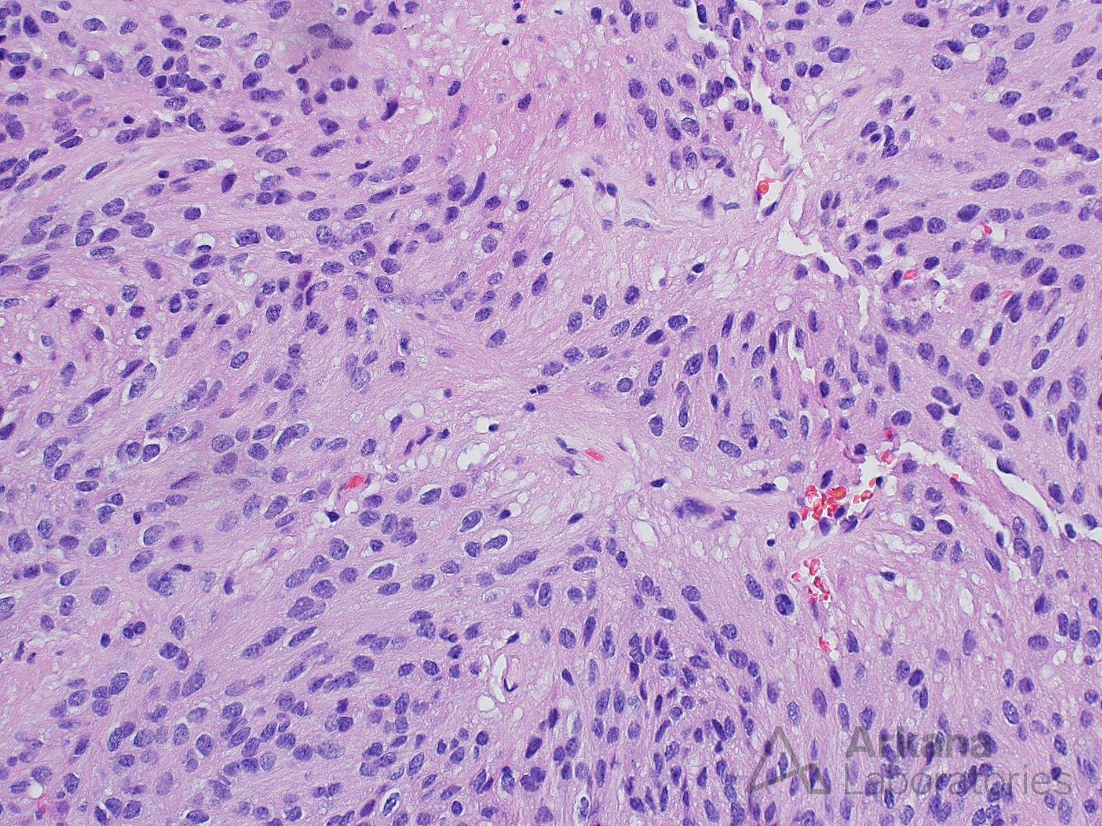
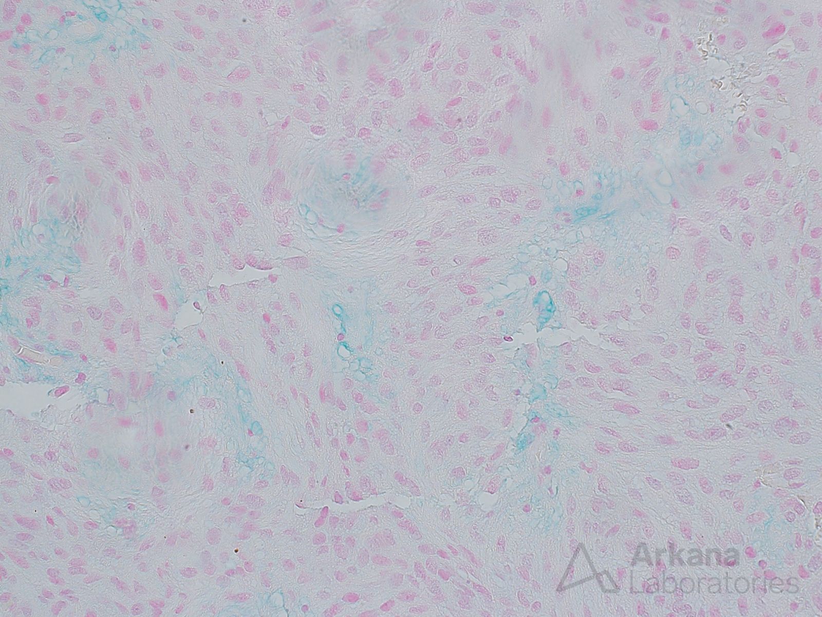
This 50-year old patient presented with a clinical history of chronic lower back pain. MRI demonstrated an intradural ovoid/sausage-shaped well-circumscribed 2.0 x 1.5 cm mass at the level of the conus medullaris that had low T1 signal, high T2 signal and diffuse contrast enhancement. The neoplastic cells were strongly GFAP positive. The proliferative fraction was relatively low.
What is your diagnosis based on the provided images of H&E and Alcian blue stained sections?
Answer:
Myxopapillary ependymoma
These are considered WHO grade 2 neoplasms.
They typically arise in the conus medullaris and filum terminale region, and represent the most common tumor at this site.
Reference(s)/Additional Readings:
- Central Nervous System Tumours: WHO Classification of Tumours, 5th Edition, Volume 6. Pages 183-185
Quick note: This post is to be used for informational purposes only and does not constitute medical or health advice. Each person should consult their own doctor with respect to matters referenced. Arkana Laboratories assumes no liability for actions taken in reliance upon the information contained herein.


