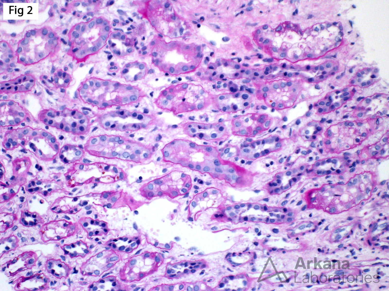
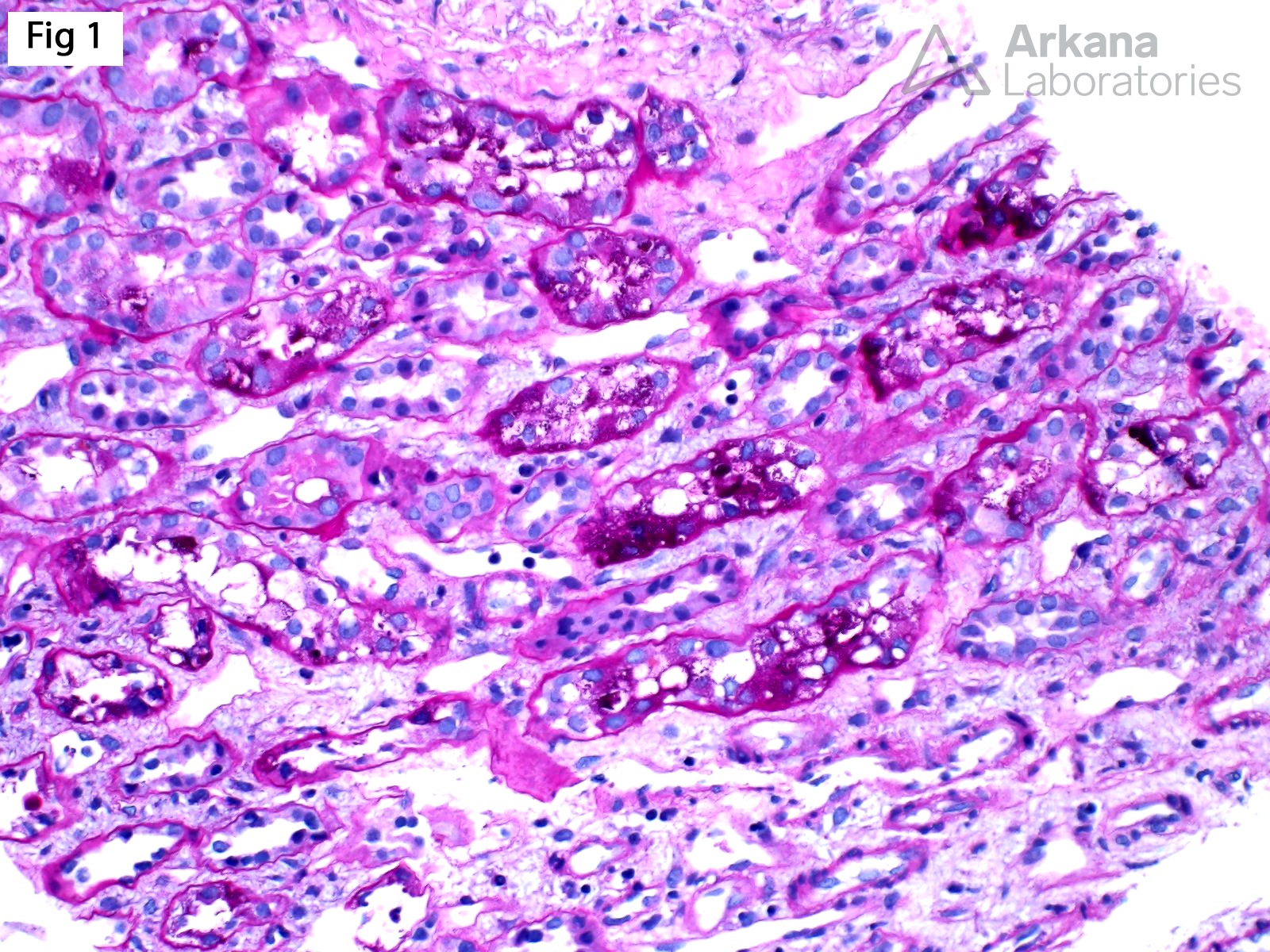
Armanni-Ebstein lesion, originally described by Luciano Armanni in 1872, is characterized by vacuolization and PAS-positive glycogen accumulation within the cytoplasm of tubular epithelial cells (Fig 1). The cytoplasm of these cells stains pale on PAS following diastase digestion (Fig 2). These changes predominantly involve the terminal straight portion of the proximal convoluted tubule and are usually seen in the outer renal medulla. Armanni-Ebstein lesions have been mostly described in poorly controlled diabetics and patients with diabetic ketoacidosis; however, similar lesions have been reported in patients with Fanconi Syndrome and alcoholic ketoacidosis. Of note, while not the original description, the term Armanni-Ebstein has also been used to describe a lesion characterized by subnuclear vacuolization without glycogen deposition, not restricted to the outer medulla or corticomedullary junction, in insulin-dependent diabetics.

References:
Zhou C, Yool AJ, Nolan J, et al. Armanni-Ebstein lesion: a need for clarification. J Forensic Sci. 2013; 58: S94-8.
Parai JL, Kodikara S, Milroy CM, et al. Alcoholism and the Armanni-Ebstein lesion. Forensic Sci Med Pathol. 2012; 8: 19-22
Quick note: This post is to be used for informational purposes only and does not constitute medical or health advice. Each person should consult their own doctor with respect to matters referenced. Arkana Laboratories assumes no liability for actions taken in reliance upon the information contained herein.


