Clinical History
This patient presented with a several year history of muscle weakness. Muscle biopsy and genetic testing were performed.
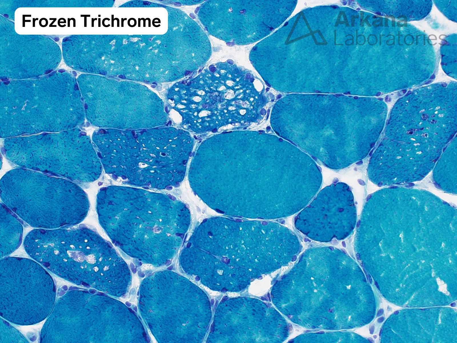
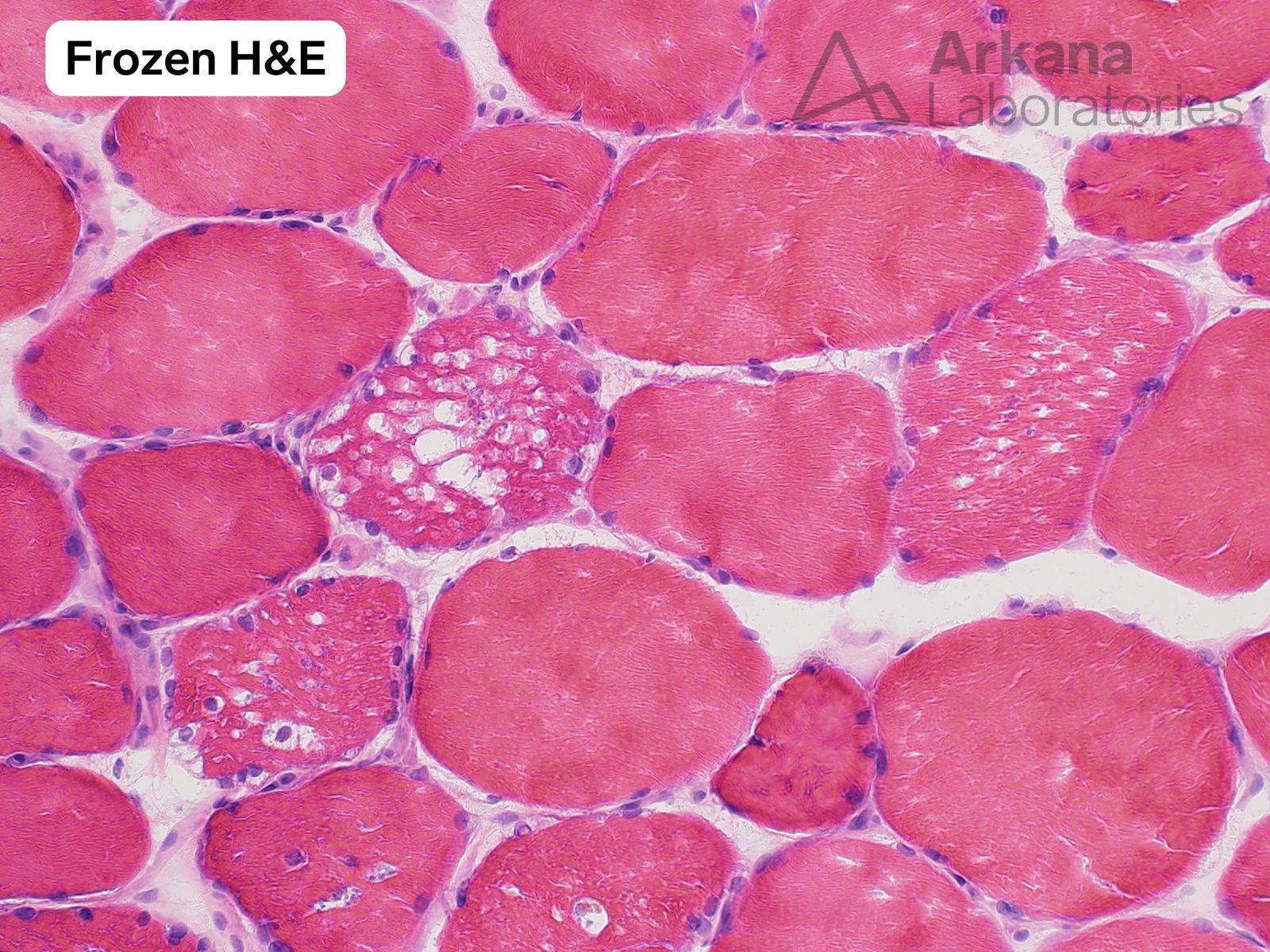
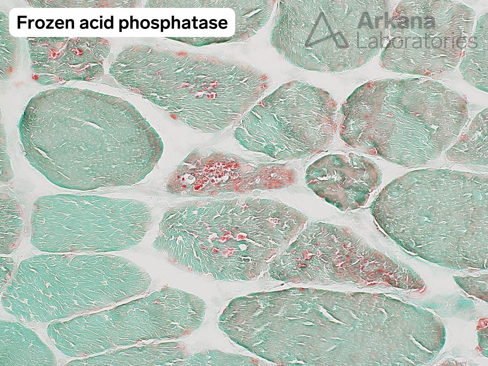
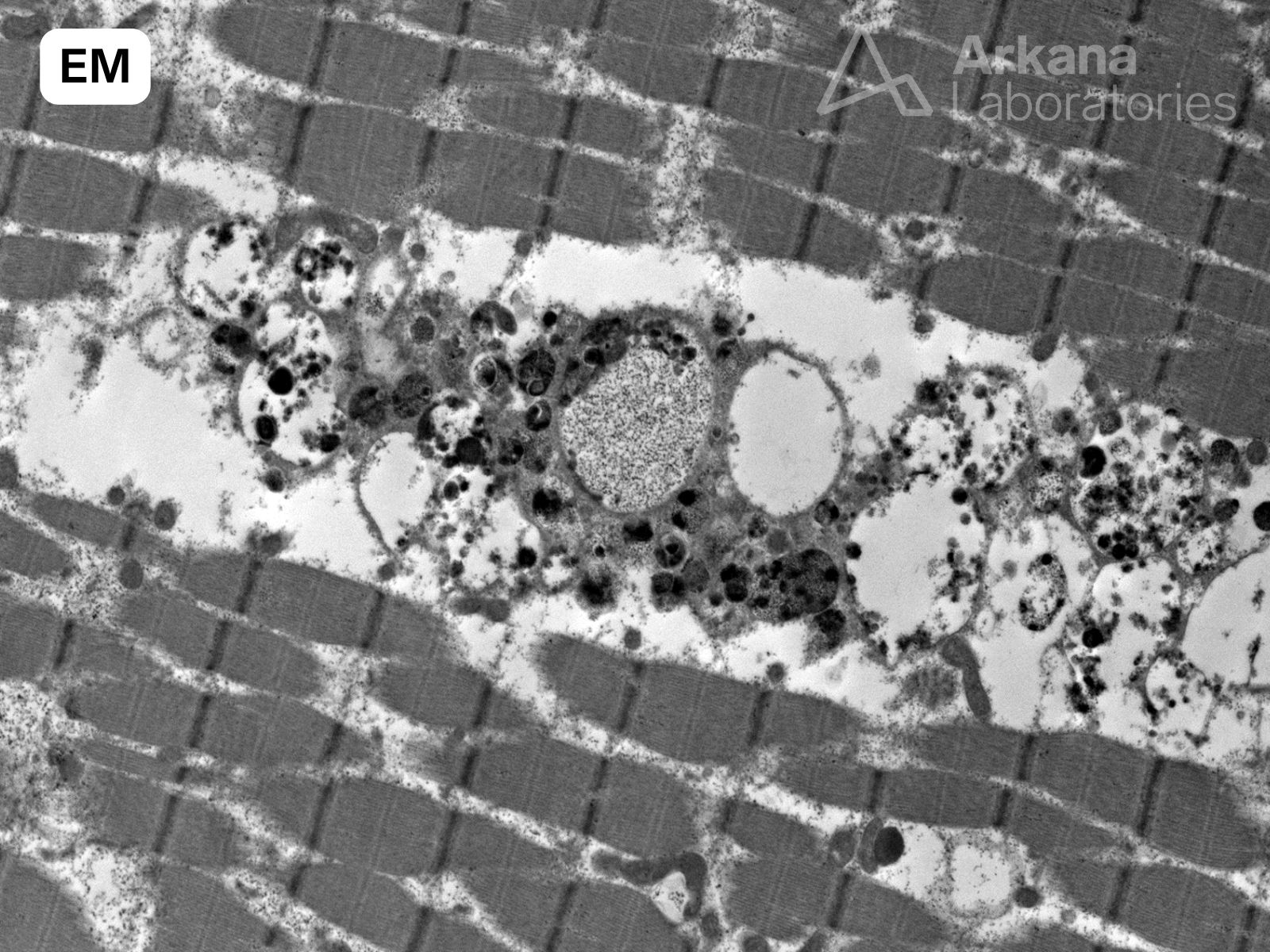
What is your diagnosis based on the provided images of H&E, modified Gomori Trichrome, acid phosphatase stained sections and electron microscopy?
Answer:
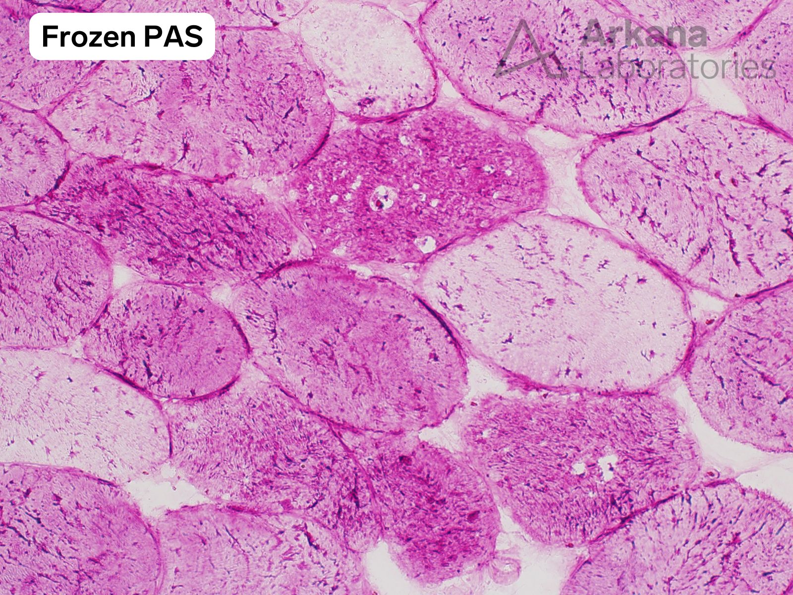
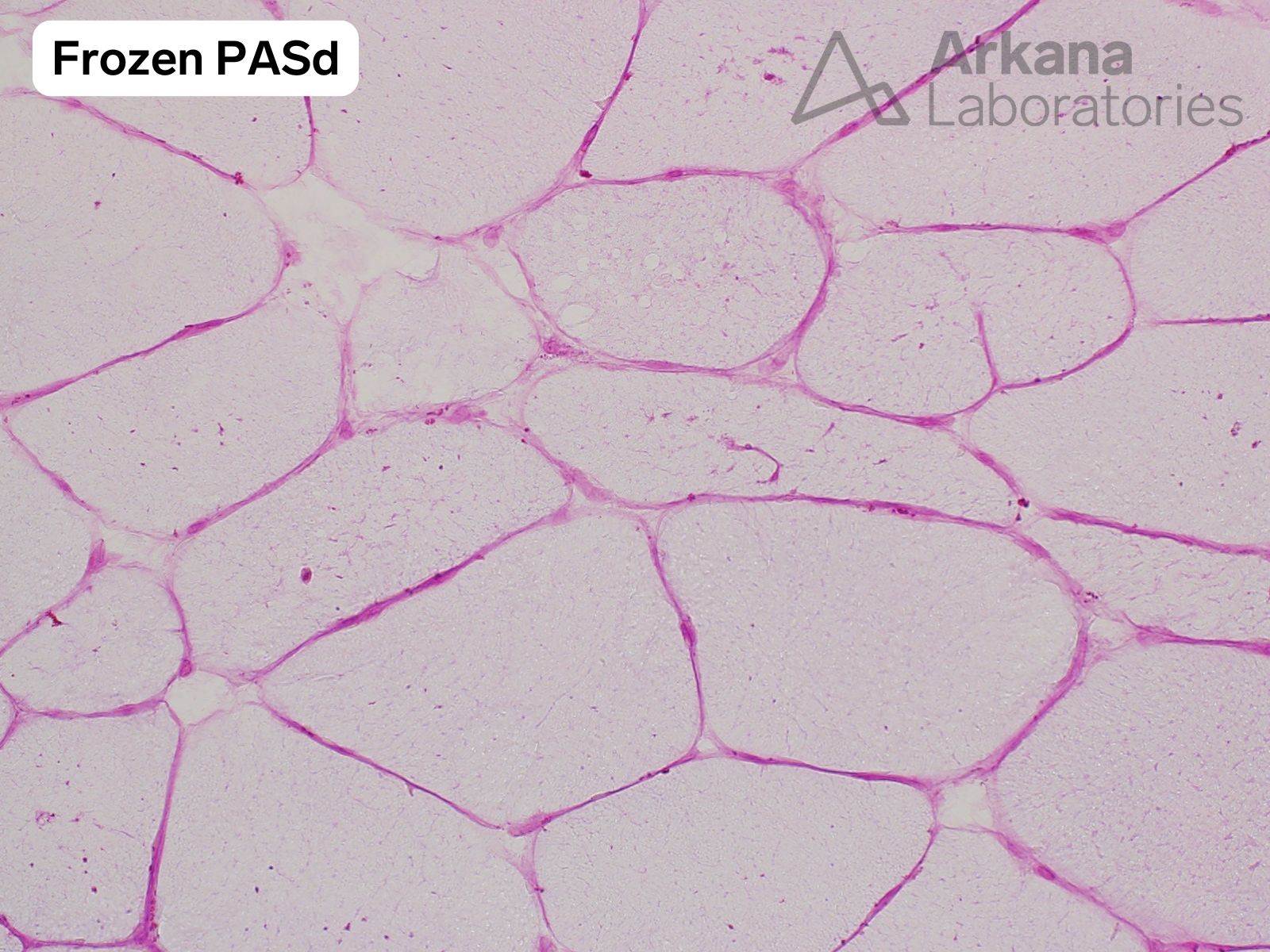
Light microscopy and electron microscopy show changes of a vacuolar myopathy with increased glycogen within lysosomes.
On discussing the pathologic findings with the patient’s physician, it was reported that genetic testing identified two different heterozygous pathogenic variants involving the GAA gene which encodes for the enzyme acid alpha-1,4-glucosidase. The patient was not being treated with hydroxychloroquine or colchicine which may cause a toxic vacuolar myopathy.
The clinical and pathologic findings are consistent with adult onset Pompe Disease (acid maltase deficiency / Glycogen Storage Disease type 2).
Reference(s) / Additional Reading:
- https://www.ncbi.nlm.nih.gov/books/NBK1261/
- Werneck LC, Lorenzoni PJ, Kay CS, Scola RH. Muscle biopsy in Pompe disease. Arq Neuropsiquiatr. 2013 May;71(5):284-9. doi: 10.1590/0004-282×20130022. PMID: 23689405.
- Taverna S, Cammarata G, Colomba P, Sciarrino S, Zizzo C, Francofonte D, Zora M, Scalia S, Brando C, Curto AL, Marsana EM, Olivieri R, Vitale S, Duro G. Pompe disease: pathogenesis, molecular genetics and diagnosis. Aging (Albany NY). 2020 Aug 3;12(15):15856-15874. doi: 10.18632/aging.103794. Epub 2020 Aug 3. PMID: 32745073; PMCID: PMC7467391.
Quick note: This post is to be used for informational purposes only and does not constitute medical or health advice. Each person should consult their own doctor with respect to matters referenced. Arkana Laboratories assumes no liability for actions taken in reliance upon the information contained herein.
