This previously healthy 30-year-old patient presented with tingling and numbness involving their extremities, followed by progressive lower extremity muscle weakness and pain. Extensive prior laboratory workup was negative for specific etiology. Electrodiagnostic studies showed evidence of an axonal distal symmetrical peripheral neuropathy.
What structure is highlighted when visualized under fluorescence microscopy?
A. Internal elastic lamina
B. Vascular amyloid
C. Micromineralization
D. Foreign material
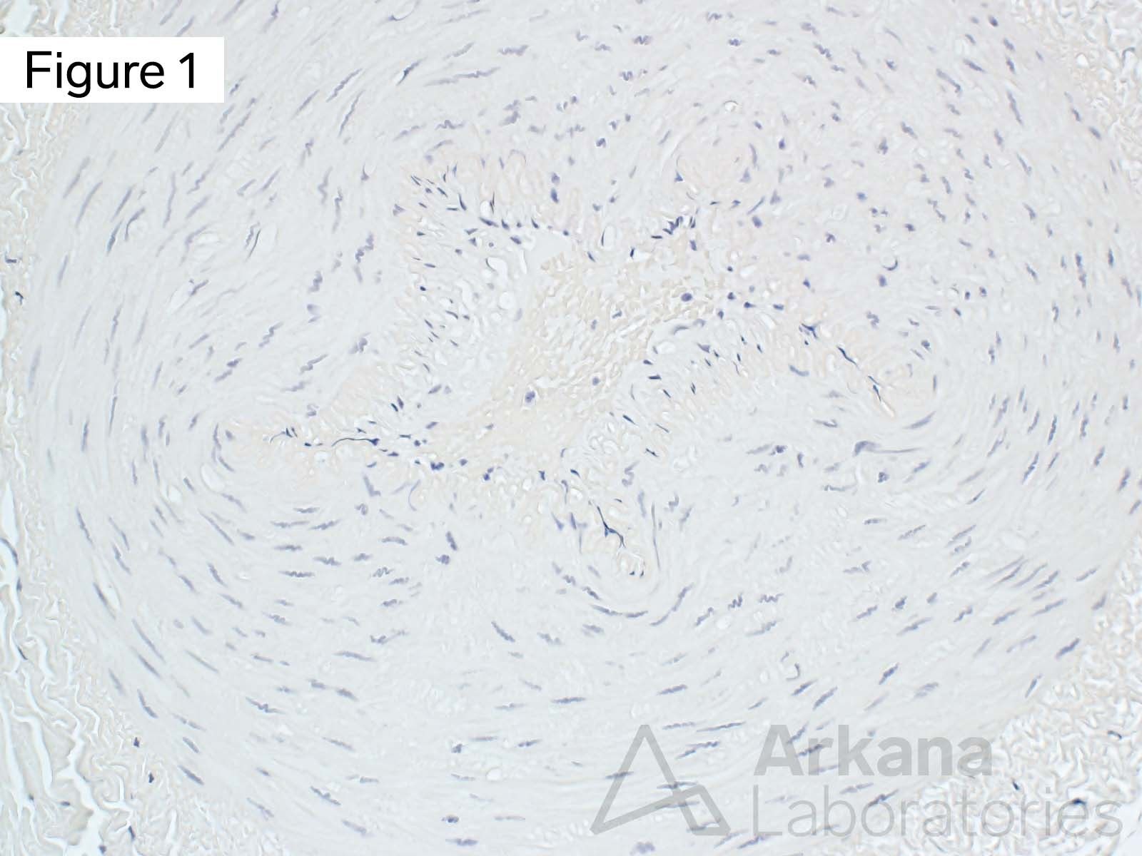
The hematoxylin counterstain in this Congo Red stained section highlights the nuclei of smooth muscle cells within this small muscular artery. The internal elastic lamina is barely visible as an undulating refractile wire-like structure. The vascular lumen contains red blood cells and a few white blood cells.
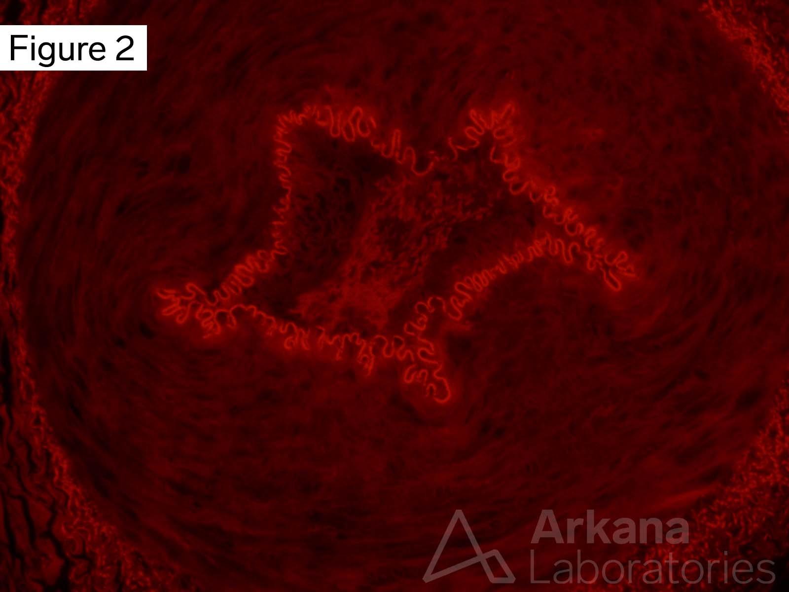
Congo red stain visualized under fluorescence microscopy using Texas Red filter nicely highlights the presence of a circumferentially intact undulating internal elastic lamina (bright red-orange). Note also some staining of the collagen bundles in the adventitia surrounding the blood vessel.
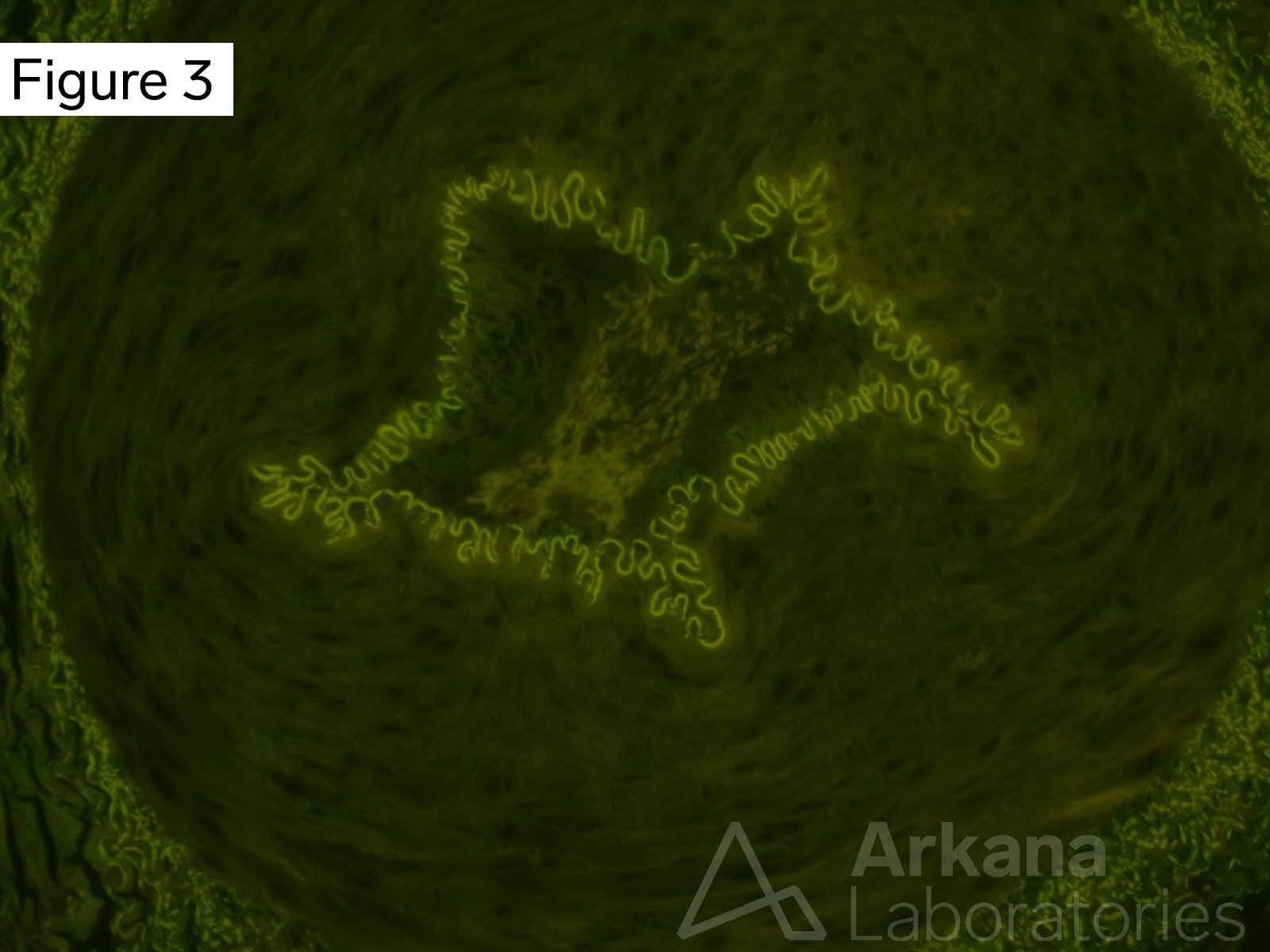
Congo red stain visualized under fluorescence microscopy using FITC (fluorescein isothocyanate) filter nicely highlights the internal elastic lamina. The tinctoral quality is white-green (as typically seen with collagen and elastin) rather than orange (characteristic of Congo Red when bound to amyloid).
Answer: Internal elastic lamina
Light microscopic examination demonstrates the presence of a small muscular artery. The internal elastic lamina is not well-visualized on bright-field microscopy but is nicely highlighted by non-specific staining for Congo Red when viewed with fluorescence microscopy using Texas Red and FITC filters.
No “salmon-orange” color on bright field (or “apple-green” birefringence on polarized light… not shown) is seen on bright-field microscopy to indicate the presence of amyloid type material. The material does not have the blue-crunchy appearance of micromineralization, and does not represent foreign material.
Quick note: This post is to be used for informational purposes only and does not constitute medical or health advice. Each person should consult their own doctor with respect to matters referenced. Arkana Laboratories assumes no liability for actions taken in reliance upon the information contained herein.