Clinical History
- This 50-year-old previously healthy patient presented with a several year history of progressive numbness, tingling and distal lower extremity muscle weakness. Electrodiagnostic studies showed features of a sensory motor axonal polyneuropathy.
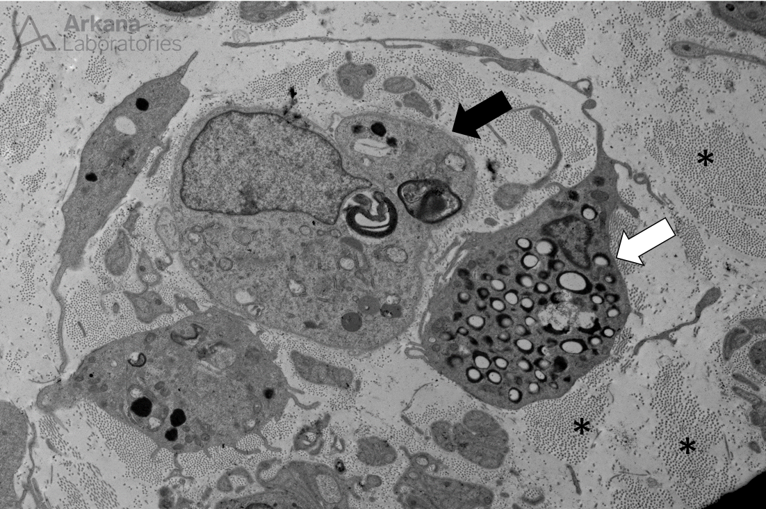
Which of the following best describes the cell indicated by the arrow in Figure #1?
A. Demyelination
B. Denervated Schwann cell
C. Onion bulb
D. Uncompacted myelin
Figure 1:
This medium power ultrastructural image shows a single Schwann cell with two small collections of myelin debris (osmophilic material) within its cytoplasm. The Schwann cell no longer has an associated axon (so-called “denervated Schwann cell”).
The adjacent cell (marked by a white arrow) is a macrophage containing myelin debris.
Note the collagen fibrils within the background endoneurium (asterix).
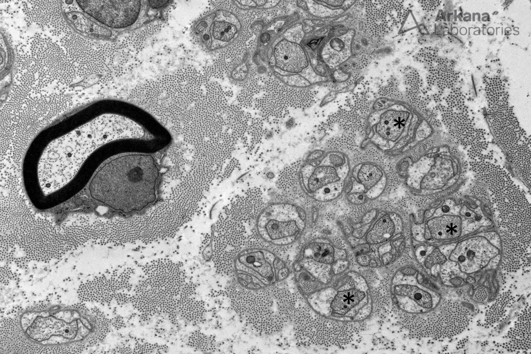
Figure 2:
For comparison, this additional medium power ultrastructural image shows:
- Myelinating Schwann cell with its associated axon (middle left half of image)
- Multiple small diameter unmyelinated axons (several marked with asterix) within small “nooks” formed by a non-myelinating Schwann cell (middle and lower right half of image)
Note: a myelinating Schwann cell will be associated with a single laxon, while an unmyelinating Schwann cell will be associated with multiple unmyelinated small diameter axons.
Correct answer: Denervated Schwann Cell
- The additional provided image shows a normal small diameter myelinated axon with its associated Schwann cell, and multiple small diameter unmyelinated axons associated with an unmyelinating Schwann cell.
Quick note: This post is to be used for informational purposes only and does not constitute medical or health advice. Each person should consult their own doctor with respect to matters referenced. Arkana Laboratories assumes no liability for actions taken in reliance upon the information contained herein.

