What is your diagnosis?

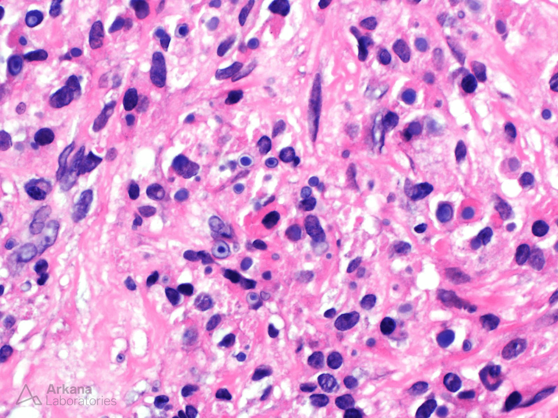
The light microscopic image depicts a renal transplant biopsy with multiple histiocytes containing intracytoplasmic, targetoid inclusions with a central core which demonstrated positive staining via a von Kossa stain. These inclusions are consistent with Michaelis-Gutmann bodies and a diagnosis of Malakoplakia.
Malakoplakia is characterized histologically by an inflammatory response typically secondary to a chronic bacterial infection. The hallmark of Malakoplakia is frequent histiocytes, sometimes showing a granular cytoplasmic appearance, with characteristic Michaelis-Gutmann bodies which develop due to decreased or ineffective lysosomal degradation of bacteria by macrophages leading to intracytoplasmic partially digested products which serve as the basis for calcium and iron deposition. The most common cause of Malakoplakia is Escherichia coli although other bacteria can lead to this entity as well (i.e. Mycobacteria, Proteus, Staphylococcus species, Rhodococcus, etc.) Our patient was known to have E. coli bacteremia serving as the nidus for the development of Malakoplakia in this case. Important to note, renal transplant recipients are a particular at risk population secondary to their altered immune status. While the urinary tract is the most commonly involved site (especially the bladder), it does occur in other organs (i.e. GI tract, lung, lymph nodes, skin, among others).
Quick note: This post is to be used for informational purposes only and does not constitute medical or health advice. Each person should consult their own doctor with respect to matters referenced. Arkana Laboratories assumes no liability for actions taken in reliance upon the information contained herein.
