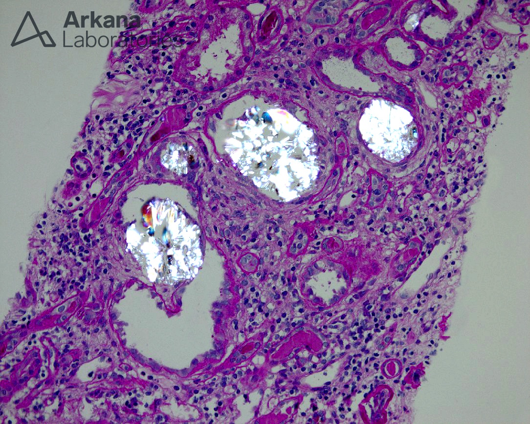
What is your diagnosis?
The light microscopic image demonstrates multiple large, polarizable intratubular crystals characteristic of calcium oxalate. While an occasional calcium oxalate crystal is not uncommon to see in renal biopsies, especially in patients with chronic kidney disease, this case demonstrated numerous crystals throughout the sample consistent with a diagnosis of renal oxalosis.
Renal oxalosis is typically a progressive tubulointerstitial disease (although it can occur acutely) with many causes which can be classified as secondary to increased ingestion, increased absorption, decreased excretion, or vitamin deficiency. Increased enteric absorption of oxalate occurs secondary to malabsorptive states such as gastric/intestinal bypass surgery (as seen in this case), prolonged antibiotic use, Crohn’s disease, pancreatic insufficiency, and short bowel syndrome. Oxalosis secondary to ingestion can occur with ingestion of ethylene glycol and oxalate containing foods such as Rhubarb, spinach, peanuts, tea, starfruit, etc. Other causes would include hypervitaminosis C, B6 deficiency, drugs (i.e. methoxyflurane, Orlistat, Praxilene, and COX-2 inhibitors), and, although less common, primary oxalosis. Treatment is aimed at removing the source of excess oxalate, a possible reversal of gastric bypass, and hemodialysis. Below are just a few references available on the causes of secondary oxalosis.
REFERENCES:
Nasr SH, D’Agati VD, Said SM, et al. Oxalate nephropathy complicating Roux-en-Y Gastric Bypass: an underrecognized cause of irreversible renal failure. Clin J Am Soc Nephrol. 2008 Nov;3(6):1676-83.
Cossey LN, et al. Oxalate nephropathy and intravenous vitamin C. Am J Kid Dis 2013; 61:1032-1035.
Quick note: This post is to be used for informational purposes only and does not constitute medical or health advice. Each person should consult their own doctor with respect to matters referenced. Arkana Laboratories assumes no liability for actions taken in reliance upon the information contained herein.

