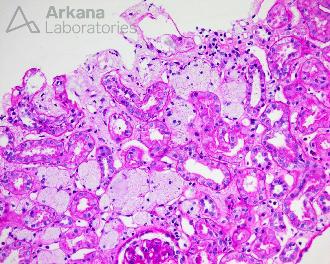
What is this finding? In a young male patient with hematuria and hearing loss, what would be your top diagnosis?
Answer: The light microscopic image depicts interstitial foam cells which, while non-specific and can be seen in longstanding proteinuria, are concerning for Alport Syndrome given the clinical scenario of hematuria and hearing loss. By electron microscopy, diagnostic findings would include glomerular basement membrane splitting, scalloping, multilamellation or “basket-weaving”, intramembranous “bread crumbs”, and abrupt transitions from areas of thin and thick GBMs. An Alport staining panel will classically show loss of staining for collagen IV alpha 5 in the majority of cases (~85%, X-linked inheritance).
Quick note: This post is to be used for informational purposes only and does not constitute medical or health advice. Each person should consult their own doctor with respect to matters referenced. Arkana Laboratories assumes no liability for actions taken in reliance upon the information contained herein.

