
This biopsy came from an elderly gentleman in his 80s, who presented with acute renal failure. His serum creatinine had increased from a baseline of 1.3 mg/dL up to 6.6 mg/dL. And in addition to that, urinalysis was positive for proteinuria and blood. His medical history included pulmonary embolism, BPH, gastrointestinal hemorrhage and GERD. Multiple serologies were ordered upon presentation, and among these, pANCA and MPO were markedly positive.
A biopsy was performed to investigate the possibility of an ANCA-mediated crescentic glomerulonephritis.
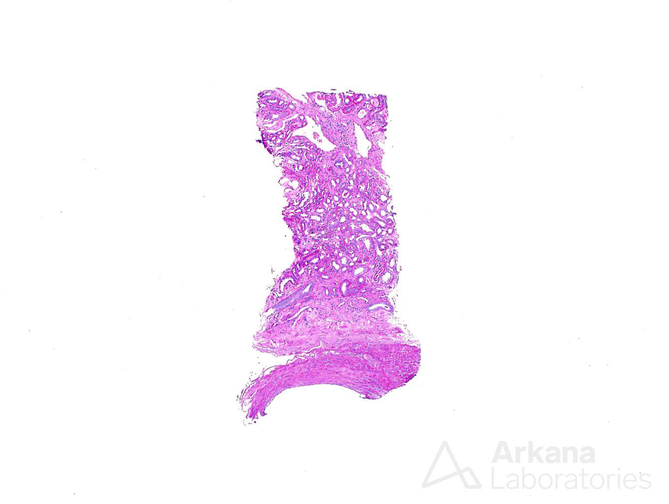
Unfortunately, the material obtained for biopsy was small in size, and the longest core, submitted to light microscopy, was only 0.4 cm. Only this minute specimen was available for evaluation on light microscopy and the tissue consisted of corticomedullary junction, without glomeruli present for evaluation.
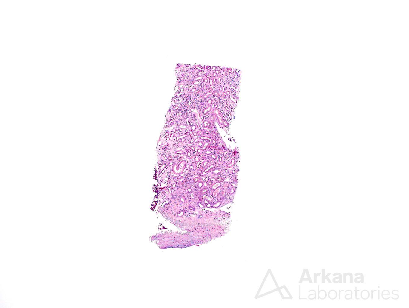
As ANCA disease manifests primarily in glomeruli and renal vessels, we proceeded to search for these elements. Thus additional, deeper sections were performed but to no avail. Although a segment of an artery was observed in the sections, still, after multiple deeper levels, no glomeruli were present on tissue submitted for light microscopy.
The tissue submitted for immunofluorescence and electron microscopy was even smaller in size than the one previously examined and we had little hope of getting to the diagnosis. The tissue on immunofluorescence contained four glomeruli, of which three were globally sclerotic. There was no evidence of immune complex deposition in the intact glomerulus. However, these results did not improve our confidence in ruling out ANCA mediated disease, and definitely did not allow us to give a diagnosis. At this point, the biopsy was shaping up to be fairly inadequate to diagnosis renal disease.
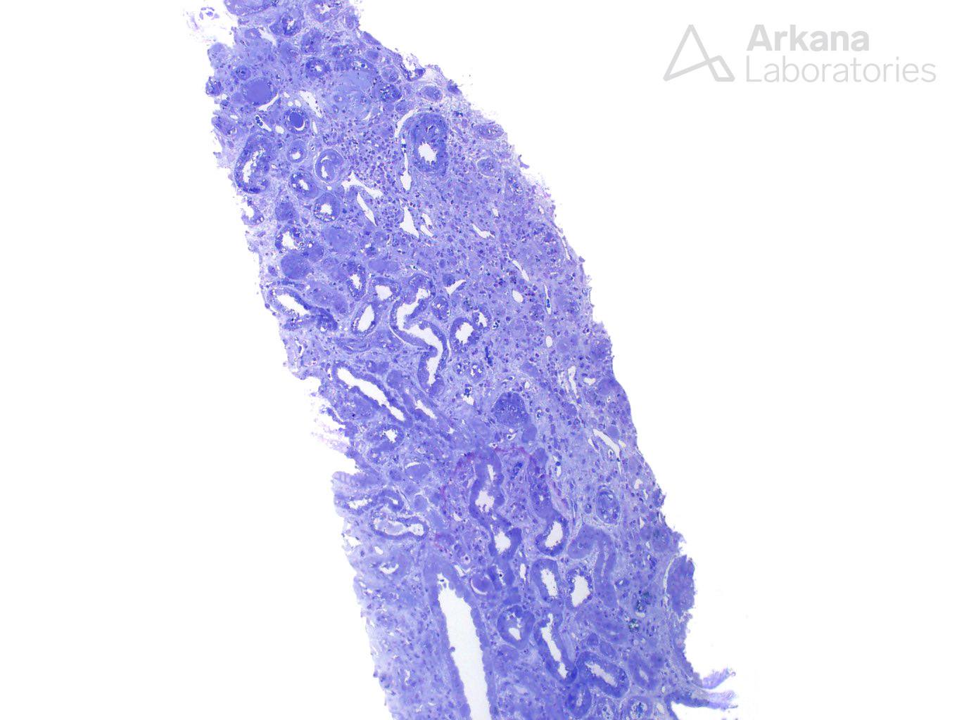
We finally recurred to our last sample – tissue submitted to electron microscopy. If one is not familiar with the procedures involved on renal biopsy, they might not know that the sample submitted to electron microscopy is always accompanied by a light microscopy counterpart. The sections described in the report as “Toluidine blue stained sections” are the ones in which the pathologist chooses which portion of the tissue, and which glomerulus, in particular, will be examined under the electron microscope. And these sections, like the other histologic stains, may carry diagnostic value if properly reviewed.
Following the pattern of the tissue examined on light microscopy and immunofluorescence, the tissue submitted to electron microscopy was a small sample. It was mostly corticomedullary junction and only one globally sclerotic glomerulus was present.
As a last resort, we decided to proceed and cut deeper toluidine blue sections. This was the last piece of tissue available for examination. On the first deeper section, one additional, intact, glomerulus was present (picture 4). This was suitable for ultrastructural evaluation, and in most instances, it would be sufficient to make us stop deeper sectioning into the block of tissue.
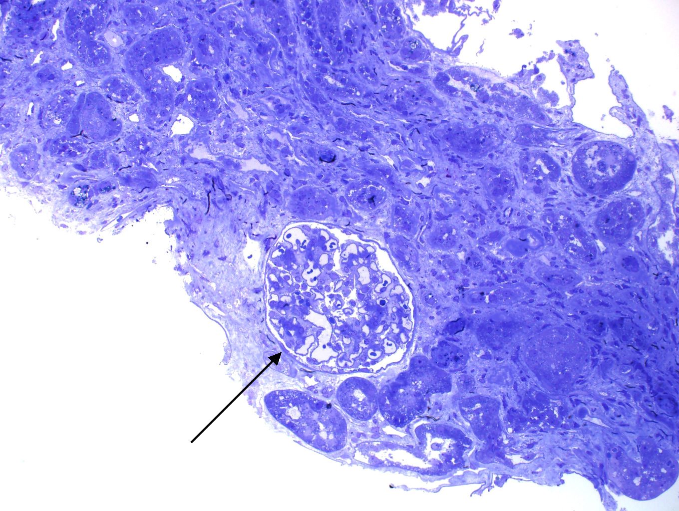
However, the question of whether an ANCA-mediated crescentic glomerulonephritis was present still lingered and not enough intact glomeruli were present to make us feel confident in excluding this possibility. This new glomerulus on deeper toluidine blue stain section served as a warning that more glomeruli could still be present in the tissue and additional deeper sections were ordered. Lo and behold, two more glomeruli were identified.
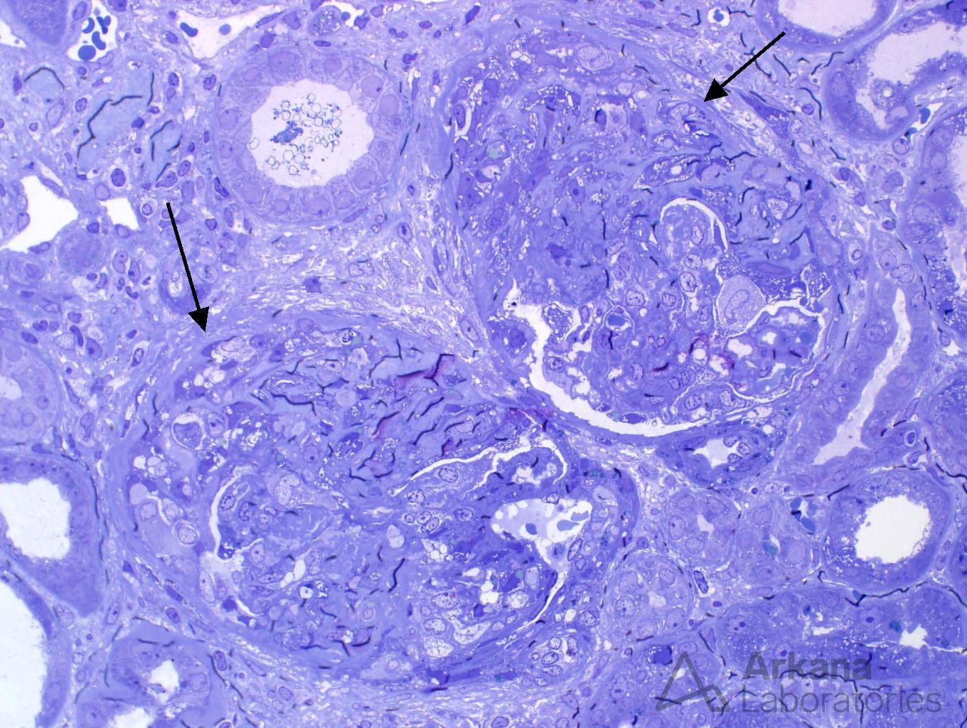
We finally had evidence on the biopsy that confirmed our nephrologist’s clinical concern and that could explain the symptomatology of our patient– two glomeruli were marked by cellular to fibrocellular crescents, with rupture of the capillary loops and were consistent with ANCA-mediated glomerulonephritis.
Through multiple steps, and multiple deeper sections, our diagnosis was finally confirmed. Digging deeper, here, there, and everywhere.
Quick note: This post is to be used for informational purposes only and does not constitute medical or health advice. Each person should consult their own doctor with respect to matters referenced. Arkana Laboratories assumes no liability for actions taken in reliance upon the information contained herein.