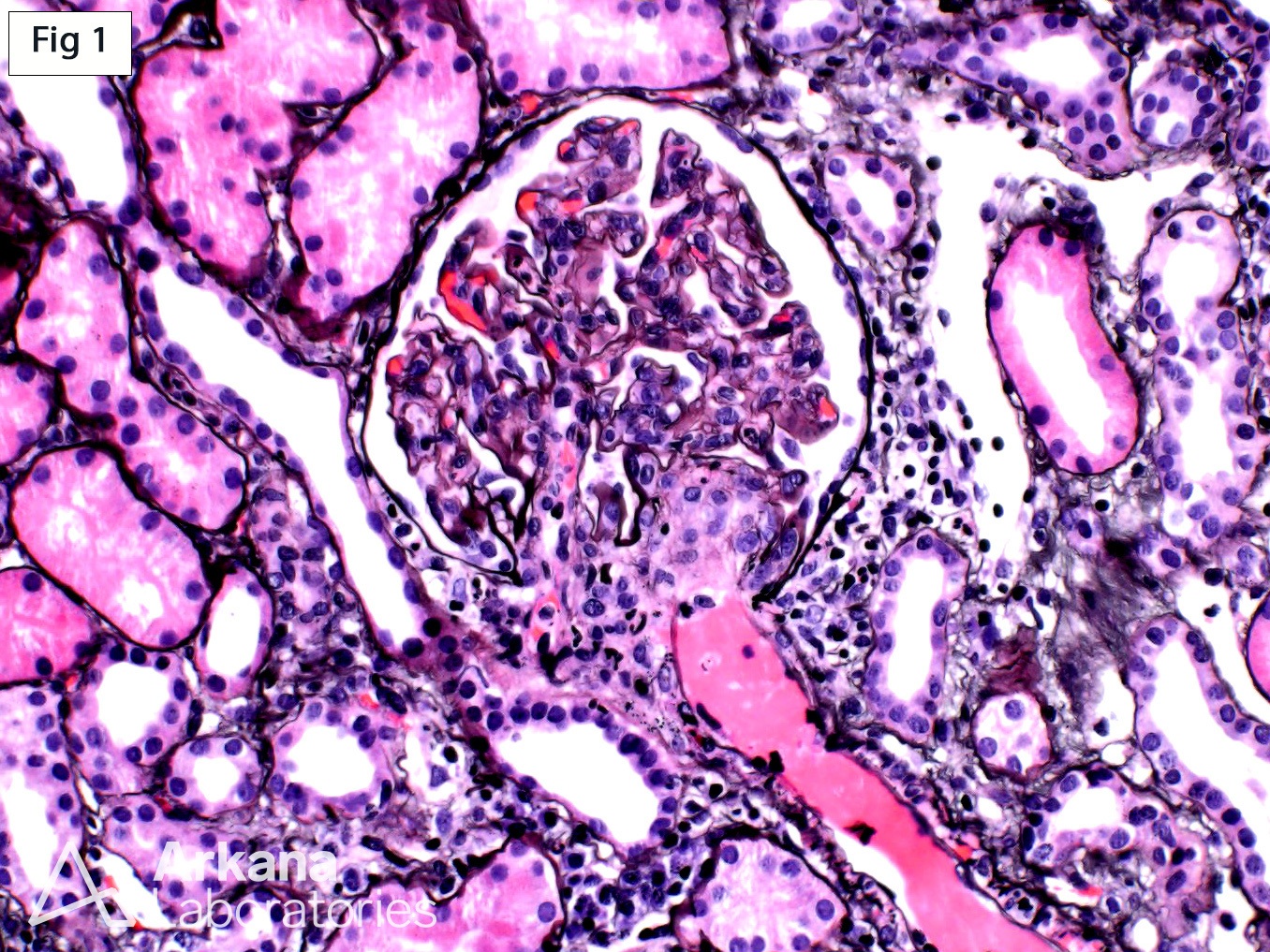
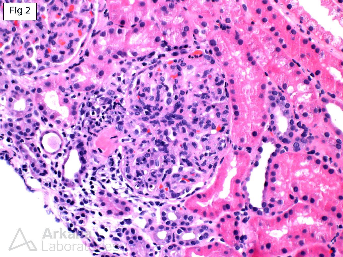
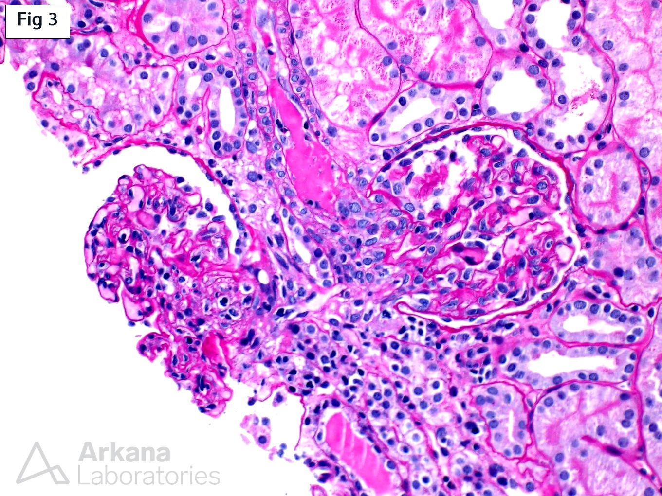
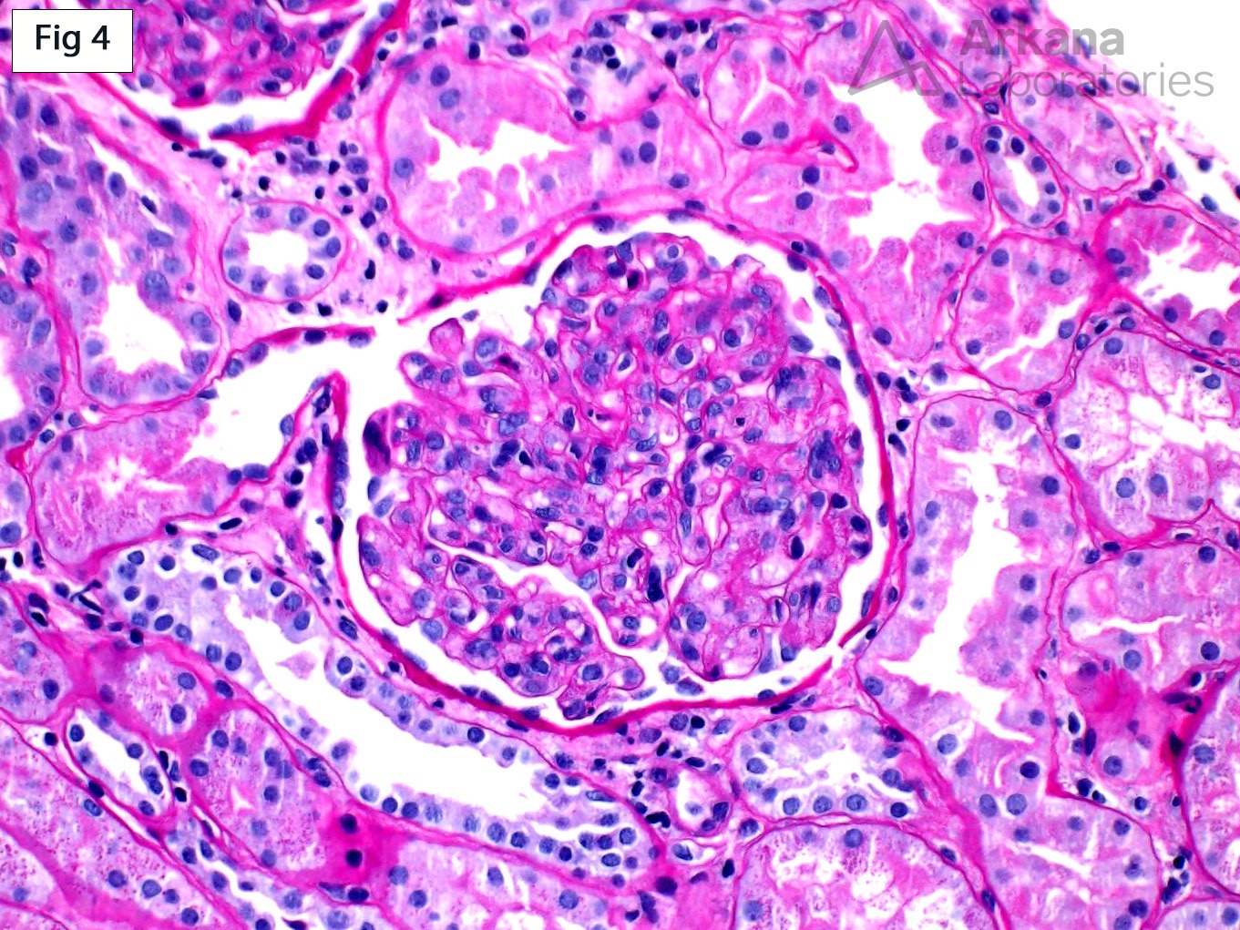
This renal biopsy performed on a 14-year-old boy shows changes diagnostic of a thrombotic microangiopathy (TMA). These changes include frequent fibrin thrombi within the arteriolar lumens and glomerular hilum, endothelial swelling, mesangiolysis and focal glomerular ischemic changes (Fig 1-3). Of note, the glomeruli frequently show marked global endocapillary hypercellularity, mostly composed of swollen endothelial cells and occasional circulating inflammatory cells (Fig 4). Immunofluorescence and electron microscopy show absence of immune complex deposition (not shown). This case illustrates that a subset of cases of TMA may show endocapillary hypercellularity and is important to avoid misdiagnosing such cases as a proliferative glomerulonephritis.
Quick note: This post is to be used for informational purposes only and does not constitute medical or health advice. Each person should consult their own doctor with respect to matters referenced. Arkana Laboratories assumes no liability for actions taken in reliance upon the information contained herein.


