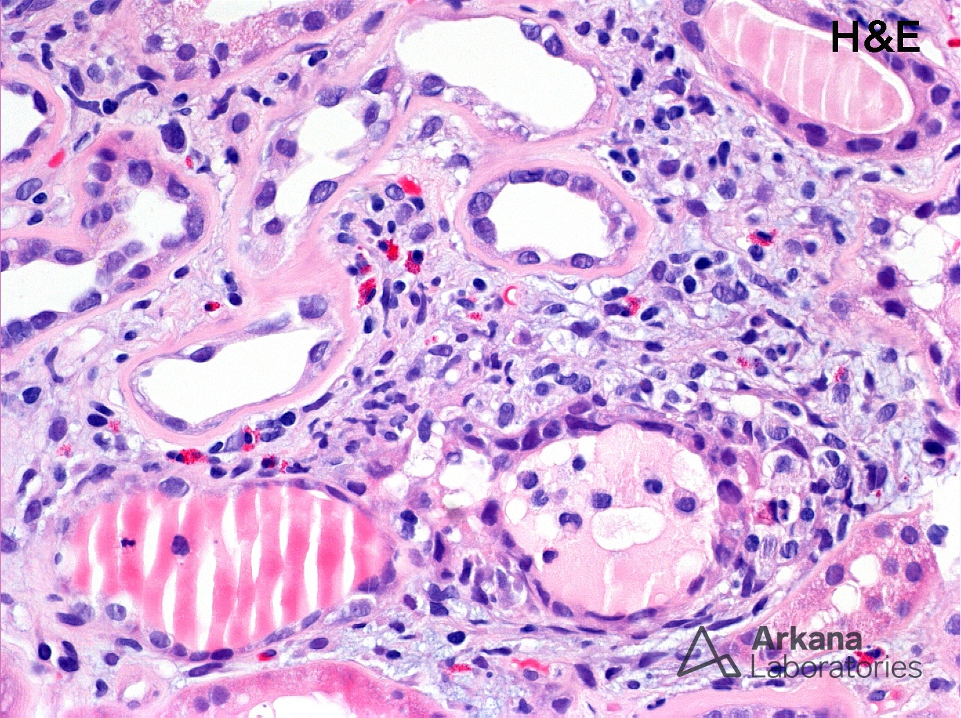
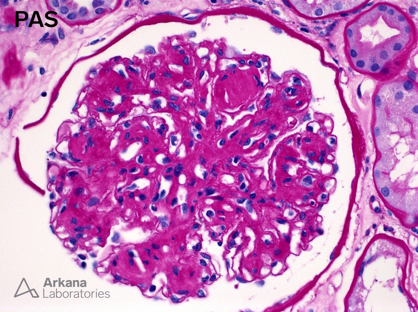
These photomicrographs are from a patient with diabetic nephropathy as evidenced by the Kimmelstiel-Wilson nodules in the glomerulus present on the PAS section. A patchy, interstitial inflammatory infiltrate rich in eosinophils is present, as seen in the H&E stained section. The infiltrate is particularly prominent in areas of fibrosis. No tubulitis is identified in the biopsy.
Interstitial inflammation rich in eosinophils is a common finding in diabetic nephropathy. When present, the pathologist is forced to determine if the inflammation warrants a diagnosis of acute interstitial nephritis. This is an important distinction because, if interstitial nephritis is present, the clinician must decide if the patient is having a drug-induced allergic reaction and if the patient should be treated with corticosteroids, which could complicate diabetic control and impose additional risk. These interstitial eosinophil aggregates in diabetic nephropathy were the focus a 2015 study by Dai, et al.1 This study found that these eosinophil aggregates are more common in diabetic nephropathy than other forms of glomerulopathy. They were associated with the interstitial fibrosis severity but, importantly, not with prescribed medications or clinical history of allergy. The authors concluded that “in diabetic nephropathy, interstitial eosinophil aggregates are… not diagnostic of an allergic interstitial nephritis”. Thus, the presence of an eosinophil-rich inflammatory infiltrate in diabetics should be interpreted with caution since this finding is often nonspecific and likely does not represent an allergic interstitial nephritis.
Quick note: This post is to be used for informational purposes only and does not constitute medical or health advice. Each person should consult their own doctor with respect to matters referenced. Arkana Laboratories assumes no liability for actions taken in reliance upon the information contained herein.


