Clinical History
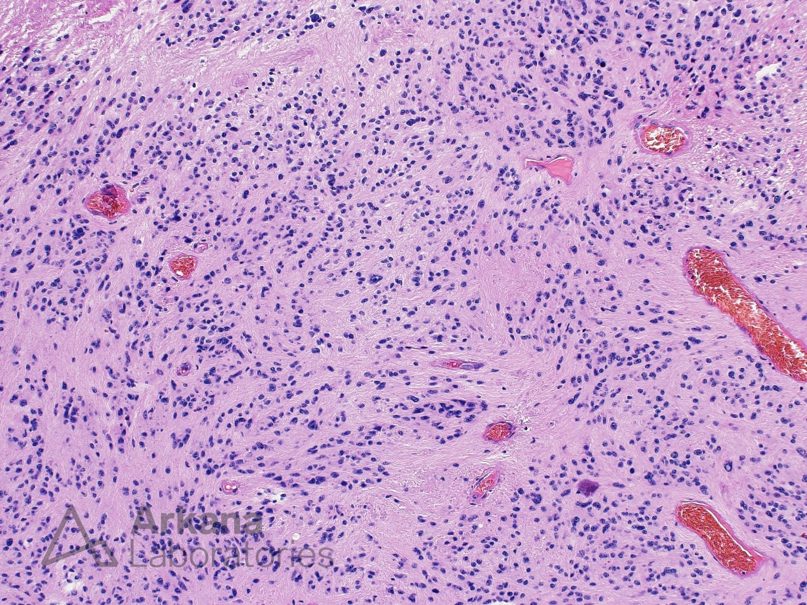
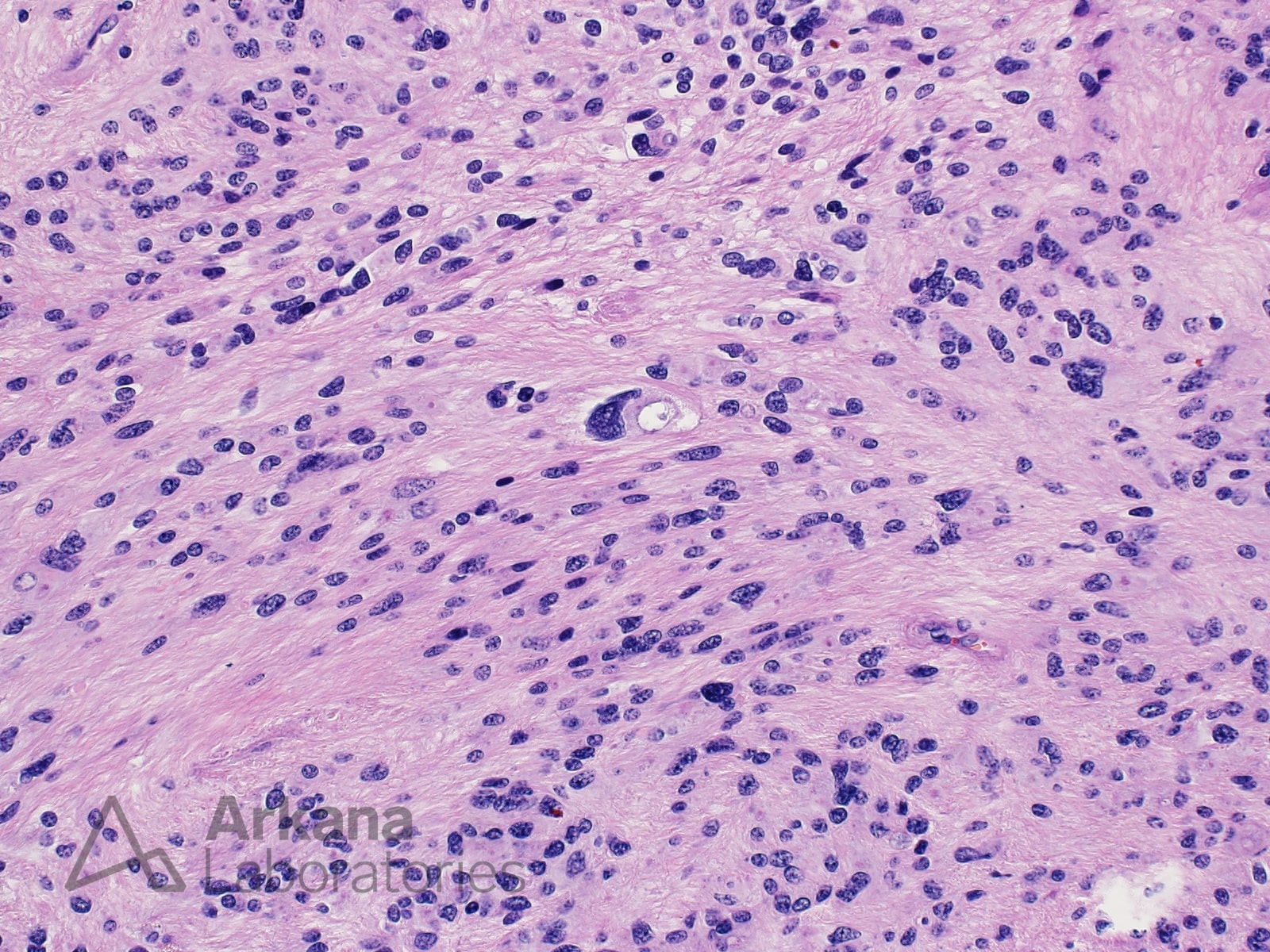
This 27-year-old patient presented with left leg numbness, low back pain and balance issues. MRI showed a 4.9 x 3.0 x 2.8 cm fourth ventricular tumor with central cystic/necrotic change. The Intraoperatively, the lesion appeared to be exophytic and was amenable to gross total resection.
The neoplastic cells showed positive staining for GFAP. Only rare tumor cell nuclei were highlighted by KI67, indicating a very low proliferative fraction.
What is your diagnosis based on the clinical history and representative pathology images?
Answer:
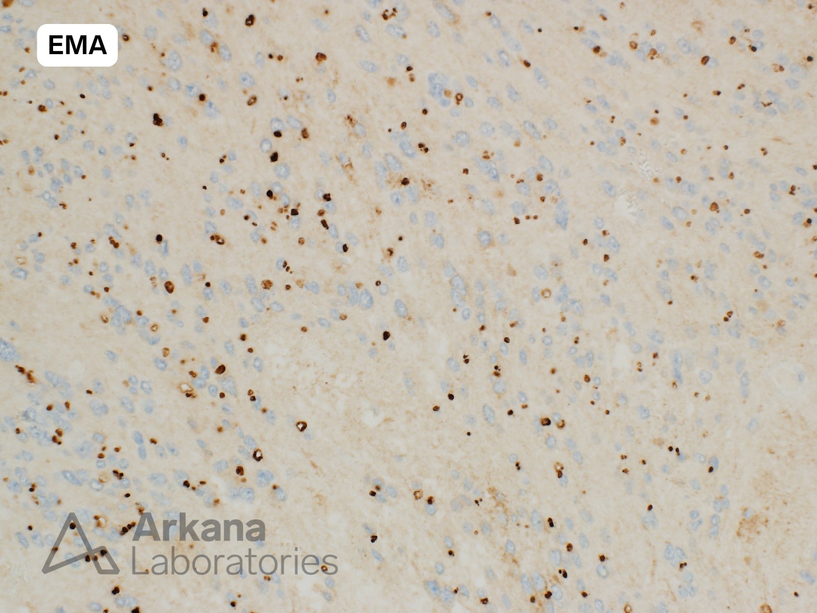
The H&E appearance and staining pattern for EMA are consistent with the diagnosis of ependymoma.
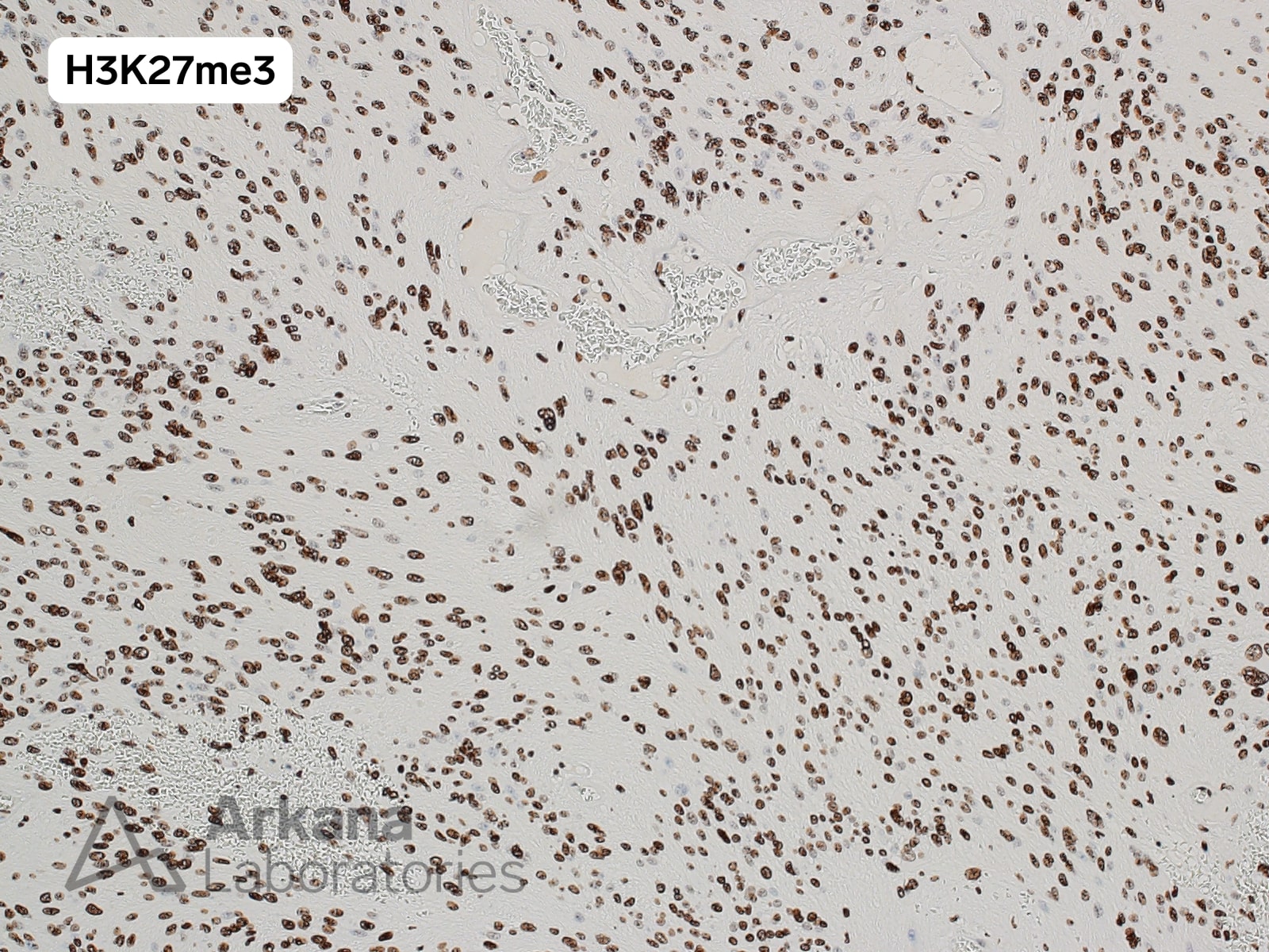
Given fourth ventricular location and retention of nuclear staining for H3K27me3, this lesion warrants classification as a posterior fossa group B (PFB) ependymoma.
References/Additional Readings:
- WHO Classification of Tumours Editorial Board. World Health Organization Classification of Tumours of the Central Nervous System. 5th ed. Lyon: International Agency for Research on Cancer; 2021. Pages 175 and 176
- Ellison DW, Aldape KD, Capper D, Fouladi M, Gilbert MR, Gilbertson RJ, Hawkins C, Merchant TE, Pajtler K, Venneti S, Louis DN. cIMPACT-NOW update 7: advancing the molecular classification of ependymal tumors. Brain Pathol. 2020 Sep;30(5):863-866. doi: 10.1111/bpa.12866. Epub 2020 Jun 23. PMID: 32502305; PMCID: PMC8018155.
Quick note: This post is to be used for informational purposes only and does not constitute medical or health advice. Each person should consult their own doctor with respect to matters referenced. Arkana Laboratories assumes no liability for actions taken in reliance upon the information contained herein.


