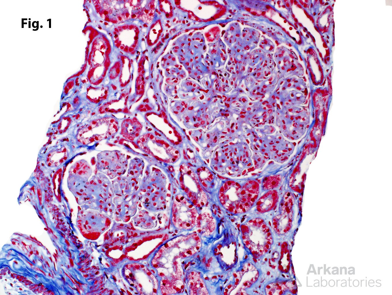
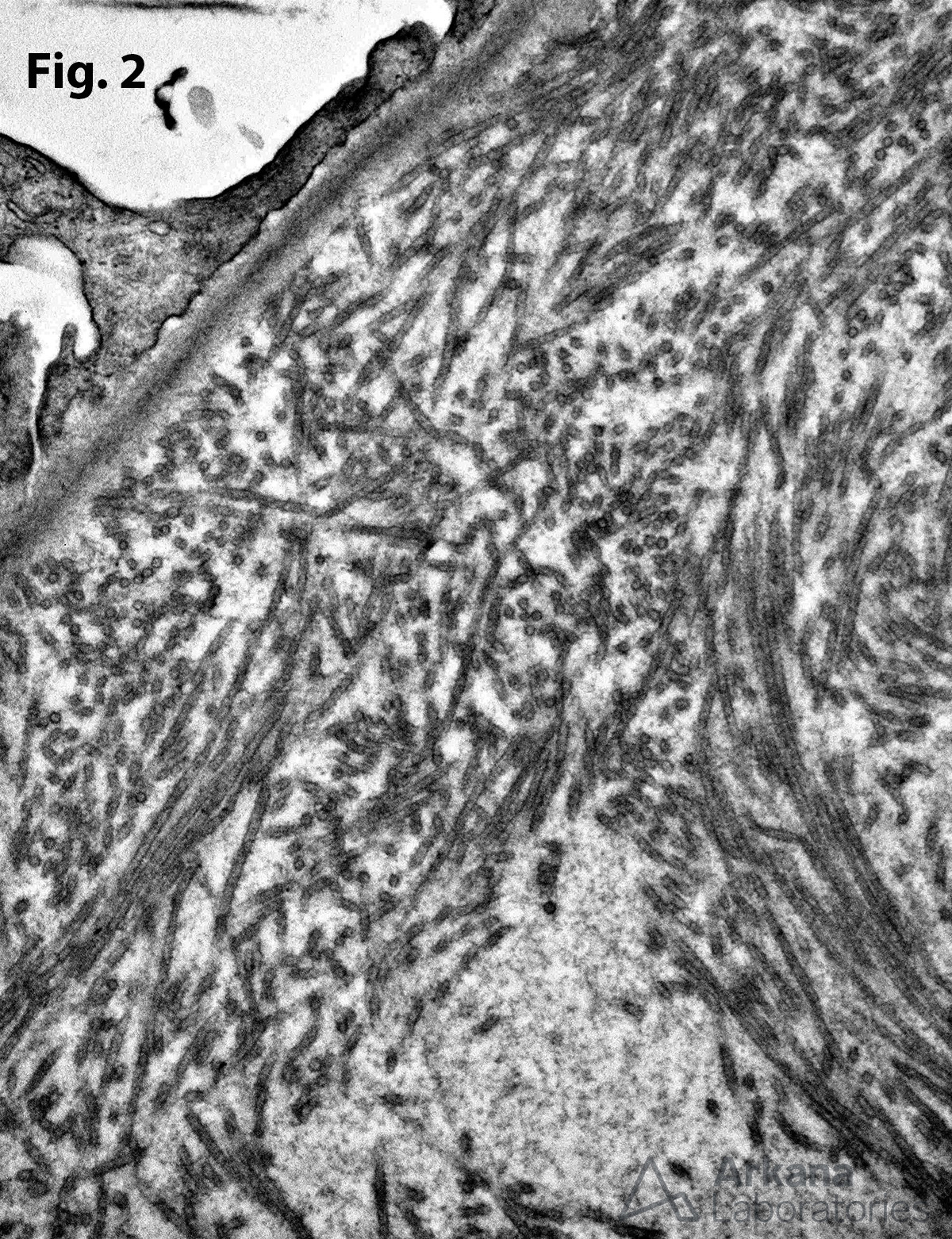
Immunotactoid glomerulopathy is one of many glomerular diseases characterized by organized deposits. Figure 1 shows characteristic glomerular changes seen with Masson trichrome staining, including capillary wall thickening, capillary proliferation, mesangial matrix expansion, glomerular tuft lobulation, and fuchsinophilic capillary wall deposits. Immunofluorescence findings may include IgG deposits and light chain restriction (kappa more often than lambda). Figure 2 shows the microtubule deposits seen by electron microscopy. These tubular type deposits characteristically show hollow centers with at least some degree of parallel arrangement. Of note, there is a strong association between immunotactoid glomerulopathy and underlying lymphoproliferative disease.
The differential diagnosis of immunotactoid glomerulopathy includes other glomerular diseases associated with organized deposits, including cryoglobulinemic glomerulonephritis, fibrillary glomerulopathy, and amyloidosis. DNAJB9 expression, which is typically seen in cases of fibrillary glomerulopathy, is not detected (see reference).
Nasr SH et al. DNAJB9 Is a Specific Immunohistochemical Marker for Fibrillary Glomerulonephritis. Kidney Int Rep. 2017 Aug 8;3(1):56-64. PMID: 29340314
Quick note: This post is to be used for informational purposes only and does not constitute medical or health advice. Each person should consult their own doctor with respect to matters referenced. Arkana Laboratories assumes no liability for actions taken in reliance upon the information contained herein.


