Clinical History:
This 50-year-old patient clinically presented with muscle weakness and elevated CPK. Myositis specific autoantibody panel was positive for NXP2 autoantibody. Muscle biopsy was performed to evaluate for myositis.
Muscle biopsy showed morphologic changes of an active and chronic inflammatory myopathy, without perifascicular atrophy (Figure #1). The FFPE sections demonstrated an incidental finding (Figures #2 – #4).
Which of the following does the incidental finding seen in Figures #2 – #4 most likely represent?
A. Traumatic neuroma
B. Neurofibroma
C. Schwannoma
D. Vasculitis
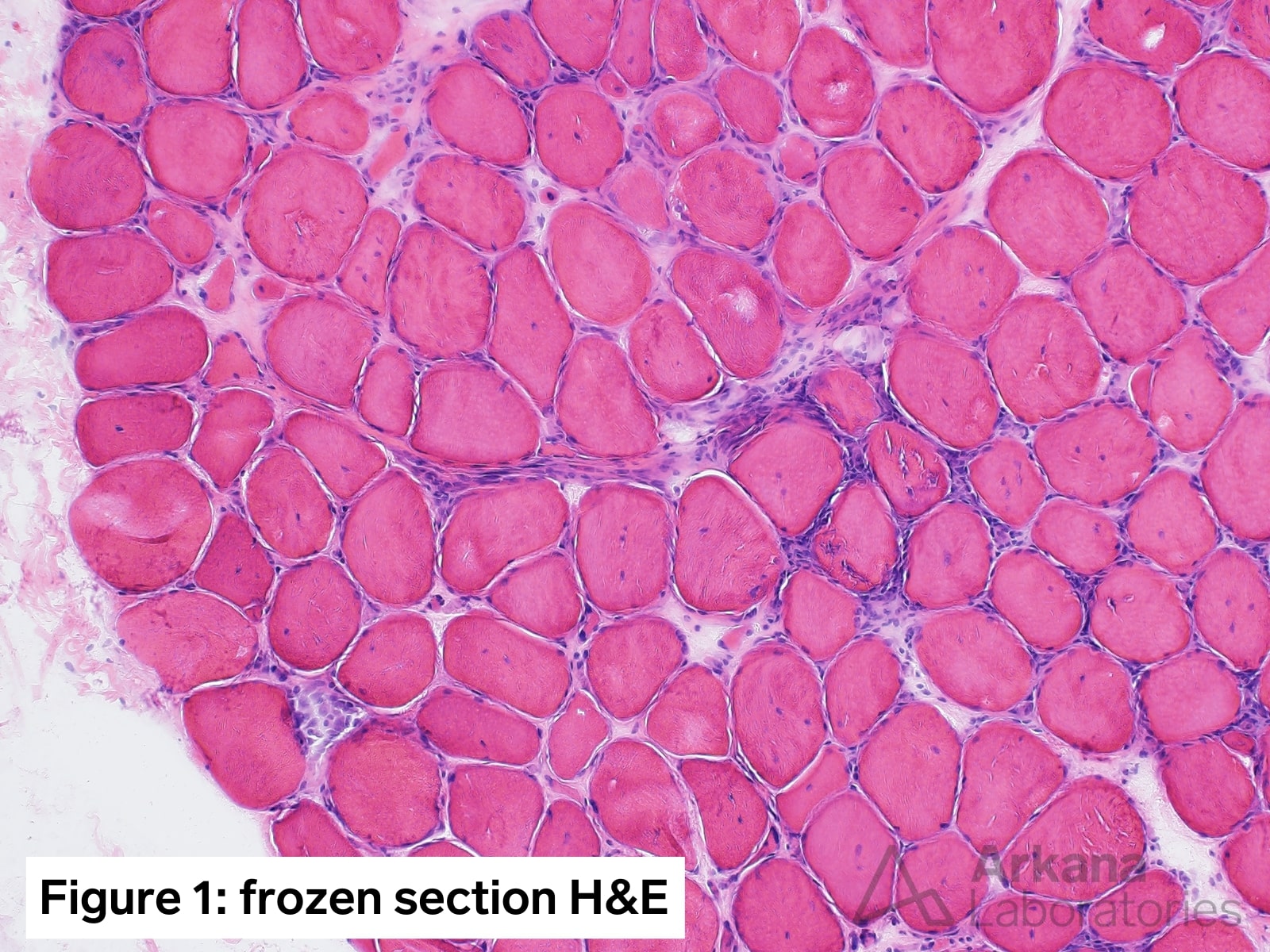
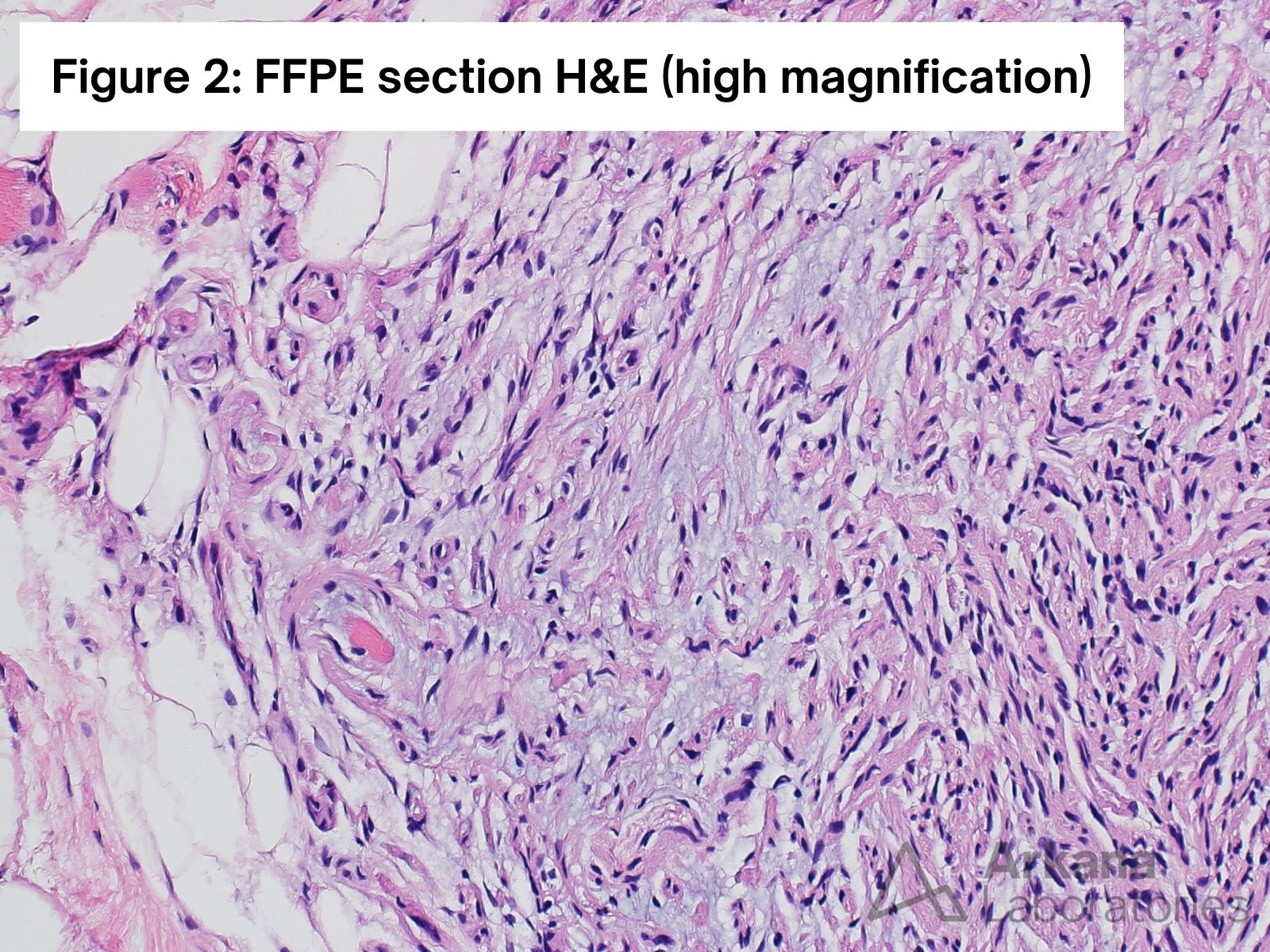
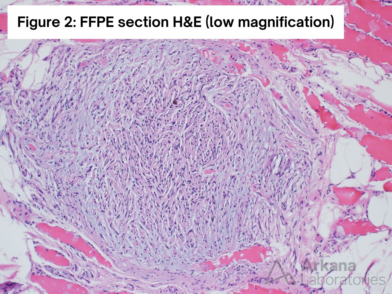
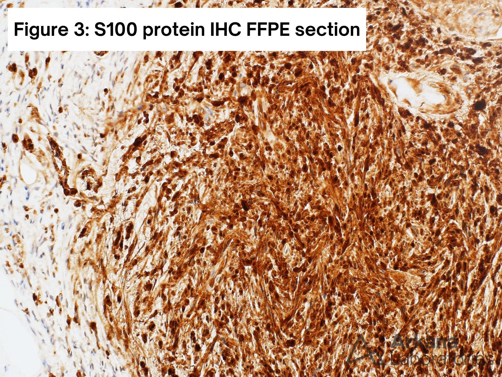
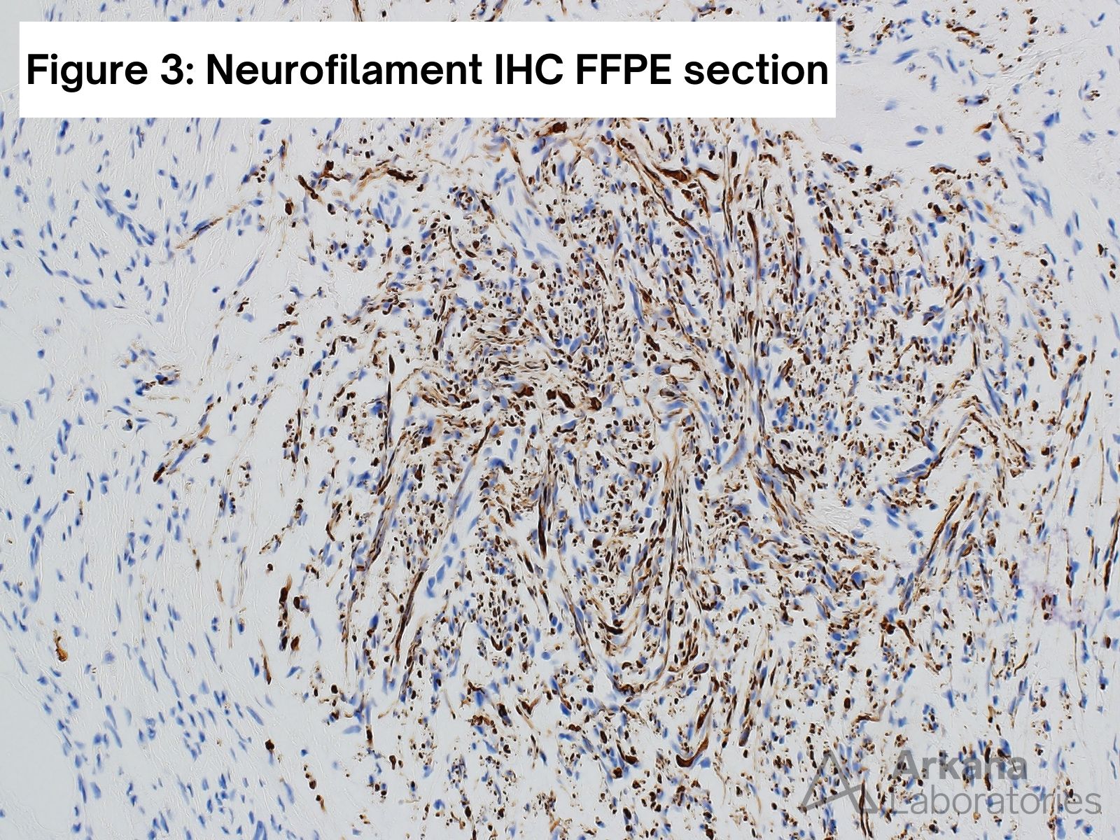
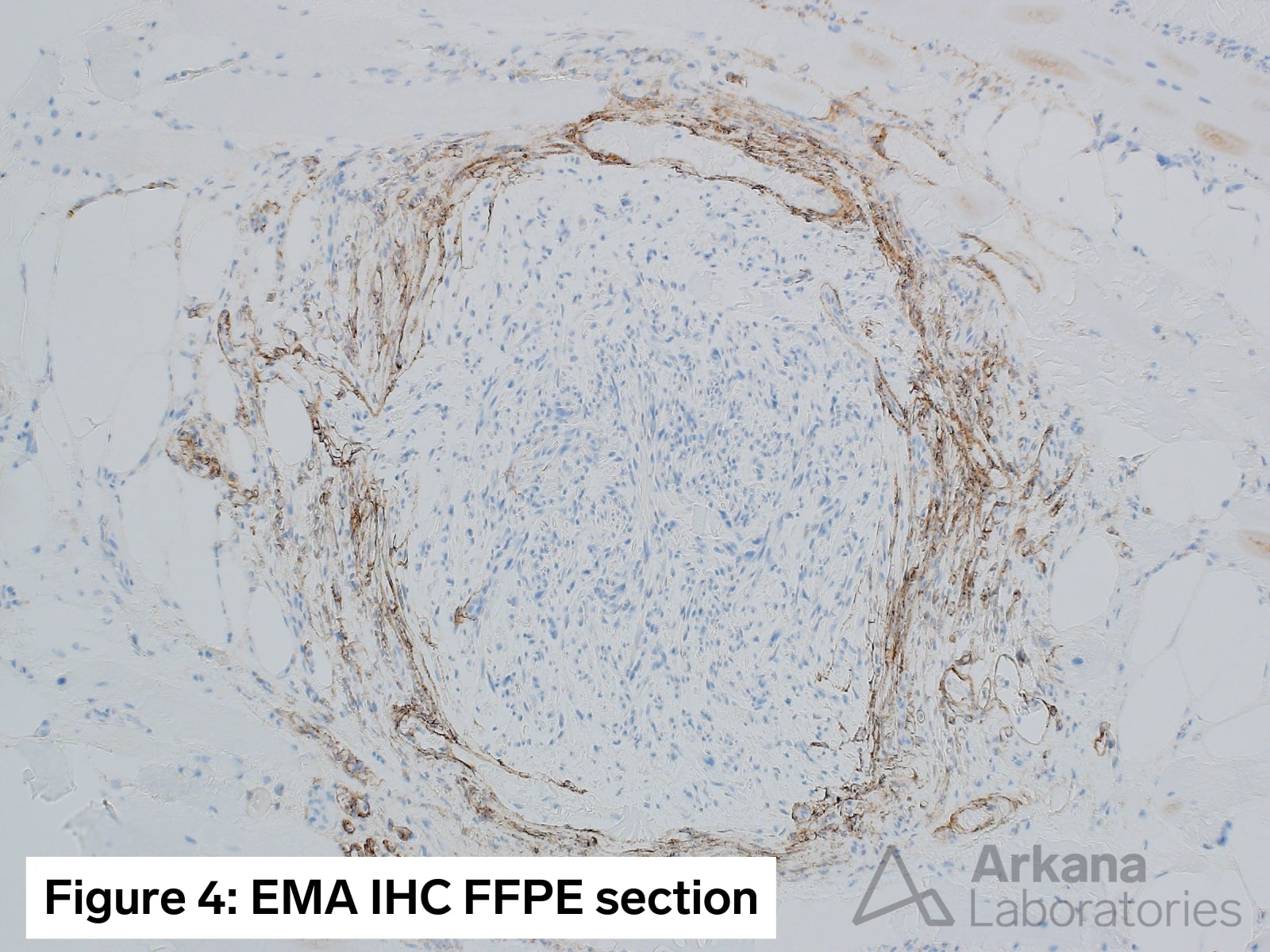
Figure 1: Medium magnification image showing skeletal muscle in cross section. Note the presence of chronic inflammation, variation in muscle fiber size, moderate to marked increase in internalized nuclei and a necrotic myofiber. Despite the reported NXP2 autoantibody (typically associated with dermatomyositis) a perifascicular pattern of myofiber injury (perifascicular atrophy) is not readily apparent.
Figure 2: Higher and low magnification images showing a somewhat circumscribed, cellular appearing spindle cell lesion with some myxoid background material. Note microfascicles toward the periphery of the lesion.
Figure 3: There is diffuse staining of the spindle cells for S100 protein (indicating that the lesion is rich in Schwann cells, while neurofilament stain highlights numerous axons arranged haphazardly within the lesion but also extending outward.
Figure 4: EMA highlights the perineurial cells. Note the somewhat ragged appearance of the perineurium and presence of perineurial cells surrounding the microfascicles toward the periphery of the lesion.
Correct Answer: A. Traumatic neuroma
The overall morphologic changes are most consistent with the presence of a traumatic neuroma.
Neurofibromas typically contain an admixture of fibroblasts, S100-positive Schwannn cells and EMA-positive perineurial cells. They tend to infiltrate the nerve (entrapped neurofilament positive axons within the substance of the lesion).
Schwannomas are typically comprised of predominantly S100-positive Schwann cells. Entrapped neurofilament-positive axons are typically sparse and toward the peripheral of the lesion.
Quick note: This post is to be used for informational purposes only and does not constitute medical or health advice. Each person should consult their own doctor with respect to matters referenced. Arkana Laboratories assumes no liability for actions taken in reliance upon the information contained herein.