Clinical History
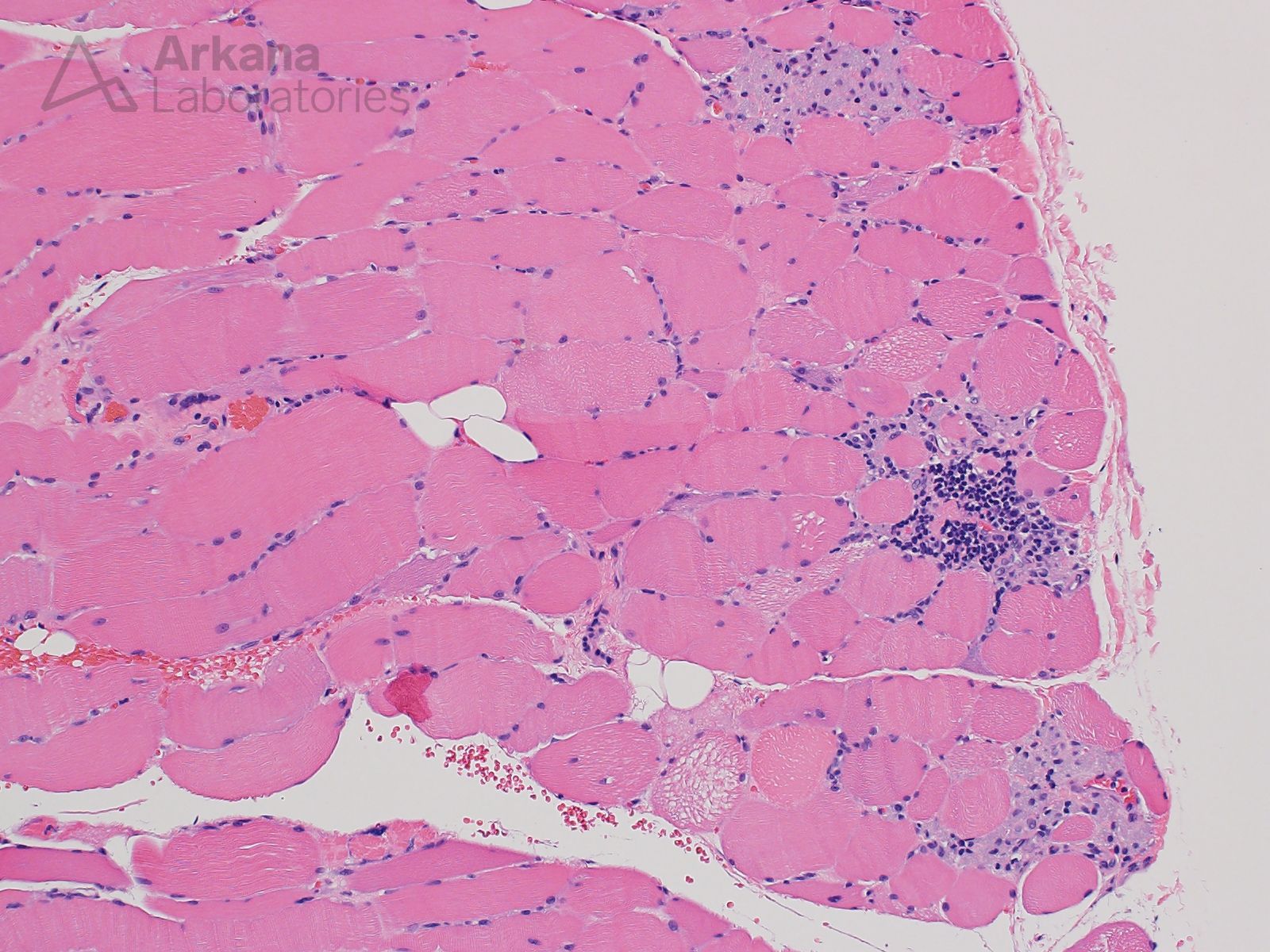
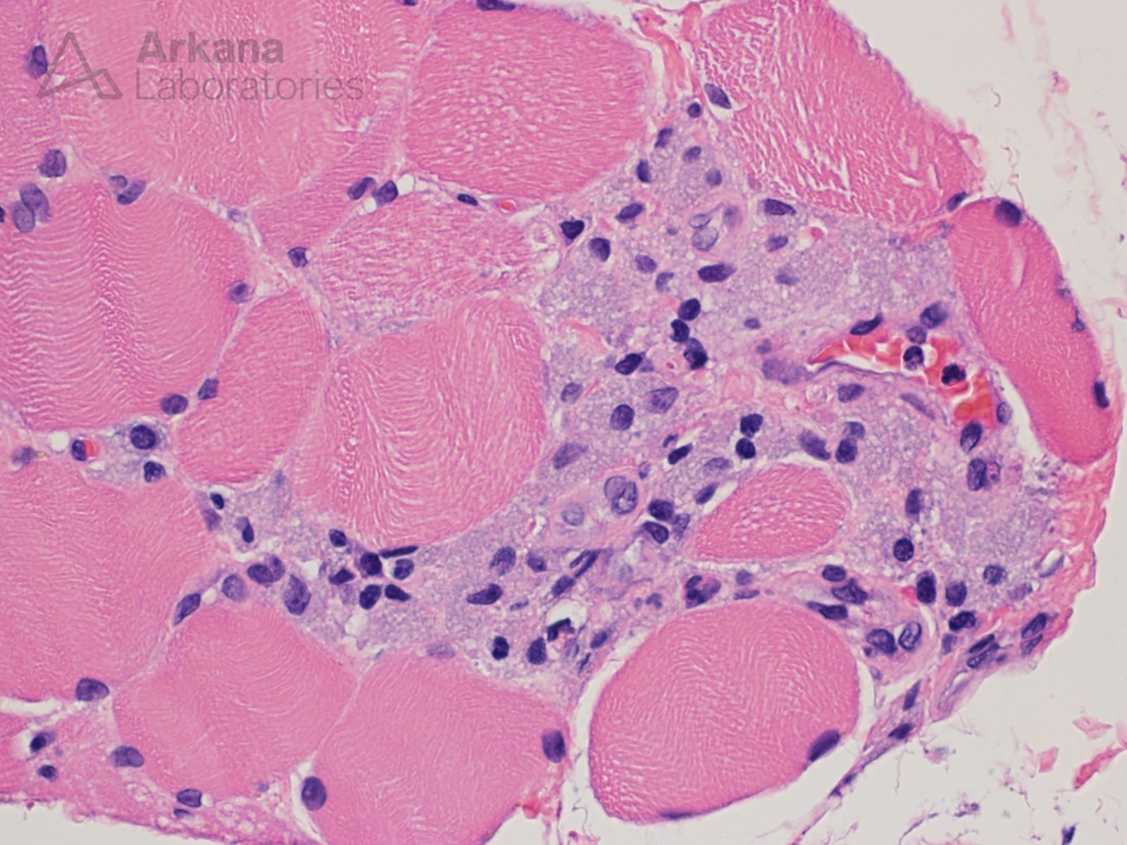
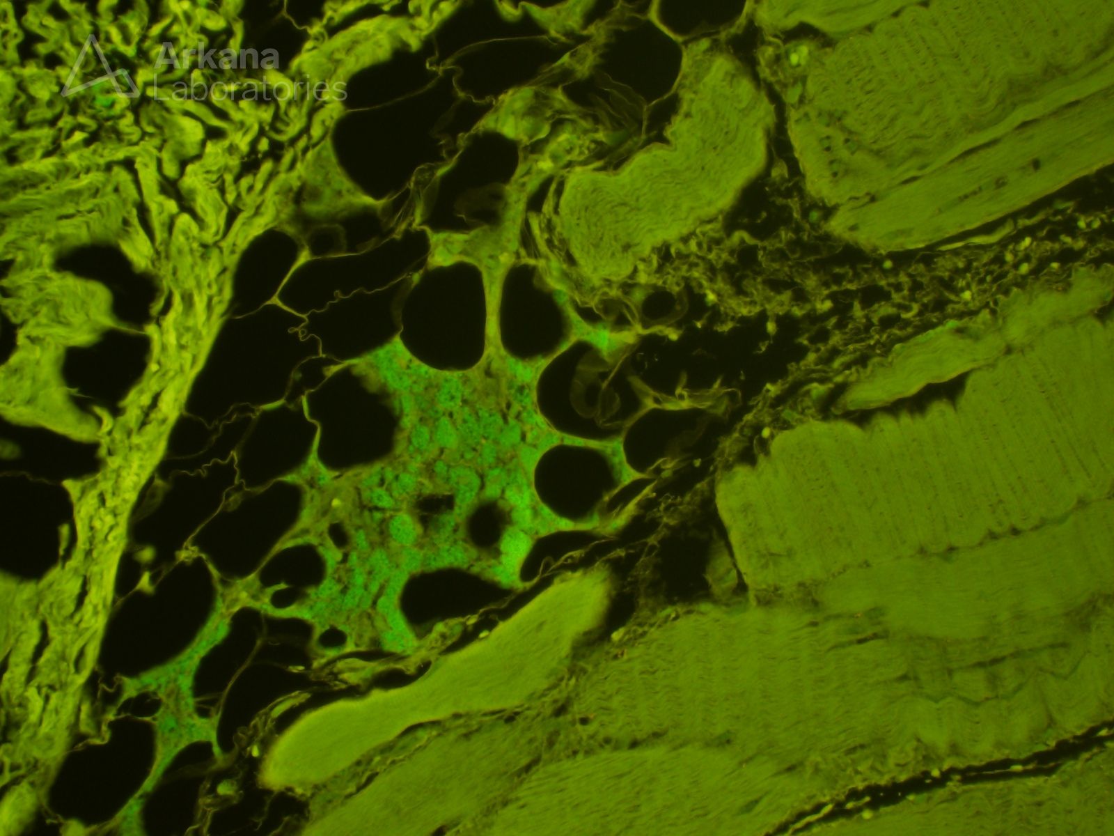
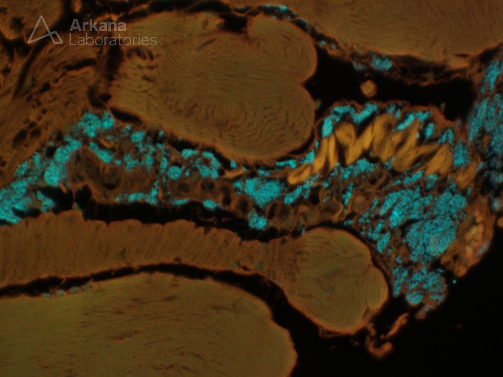
This 60-year old patient presented with a several month history of progressive upper and lower extremity muscle weakness with associated significant elevation of CPK. Muscle biopsy showed morphologic changes of an active inflammatory myopathy. Hematoxylin and eosin stained sections also showed additional findings that prompted the performance of a special stain that is evaluated under fluorescence microscopy.
What do the findings on the below images indicate?
Answer:
These additional findings are consistent with prior vaccination at the site of this patient’s muscle biopsy. Morin stain demonstrates the presence of “alum” (aluminum vaccine adjuvant) within macrophages which is characteristic of “macrophagic myofasciitis.”
Reference/Additional Reading:
- Chkheidze R, Burns DK, White CL, Castro D, Fuller J, Cai C. Morin Stain Detects Aluminum-Containing Macrophages in Macrophagic Myofasciitis and Vaccination Granuloma With High Sensitivity and Specificity. J Neuropathol Exp Neurol. 2017 Apr 1;76(4):323-331. doi: 10.1093/jnen/nlx011. PMID: 28340105; PMCID: PMC5901095.
Quick note: This post is to be used for informational purposes only and does not constitute medical or health advice. Each person should consult their own doctor with respect to matters referenced. Arkana Laboratories assumes no liability for actions taken in reliance upon the information contained herein.