MIC Current Offerings:
- NIH-funded projects
- Industry-sponsored projects
- Contract research services for multiplex imaging and image processing
From glass to digital, from data to actionable analysis interpretation - the Multiplex Imaging Center is focused on applying novel imaging technologies to advance understanding of disease.

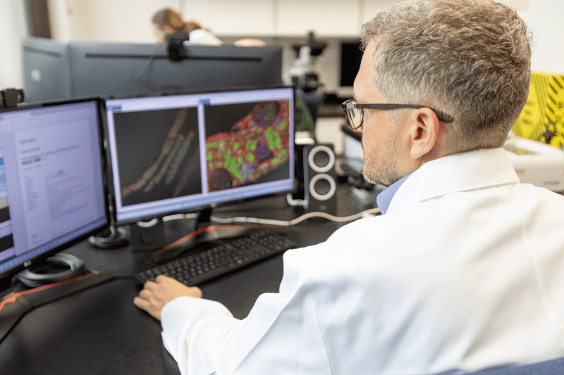
Founded and directed by Dr. Shana Coley, M.D., Ph.D., the Multiplex Imaging Center (MIC) is supported by an imaging research scientist and an imaging data scientist. This Arkana core research facility is located in the same building as Arkana’s clinical lab, in close proximity to both renal pathology and neuropathology expertise.
The MIC serves multiple research projects at Arkana, both NIH-funded projects and industry-sponsored projects, as well as internal R&D. We also offer contract research services for multiplex imaging and image processing.
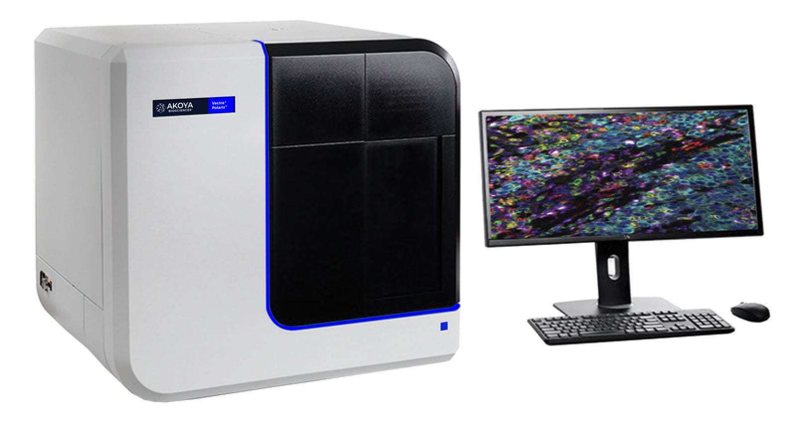
We are a certified partner with Akoya Biosciences in their CRO Partnership Program, specializing in multiplex imaging of human tissues. We have an Akoya Polaris 9-color fluorescence and brightfield whole slide scanning microscope with InForm software for spectral unmixing, Halo AI image processing software, Halo Link access for remote collaborations, and specialized modules for niche image processing projects. The MIC also has access to an Olympus VS200 high-throughput fluorescence and brightfield scanning microscope, and a Leica laser scanning confocal microscope located on the same corridor, for both high-volume and high-resolution advanced microscopy.

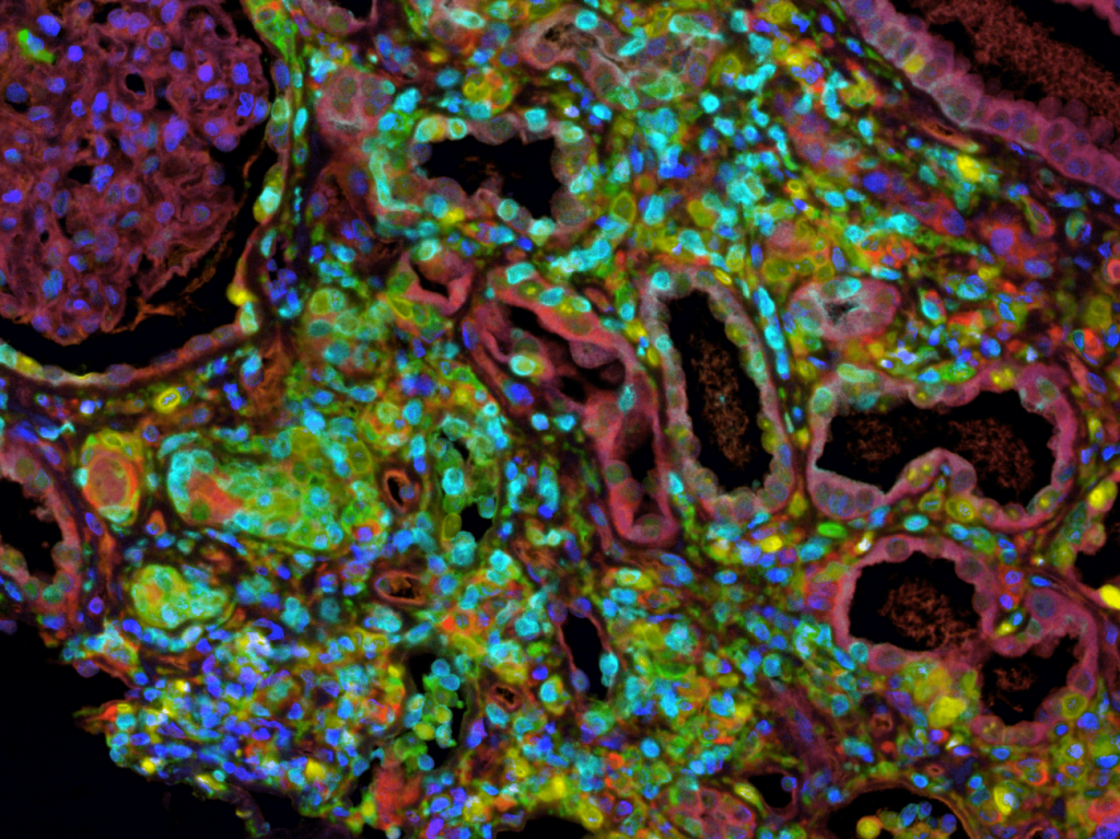
Multiplexed immunofluorescent staining panels are developed for human tissues using Opal TSA technology and the Leica Bond RX autostainer. Whole slide digital images of these tissues are generated using our Akoya Phenoimager HT, with spectral unmixing using InForm software. Digital images are processed using Halo AI software from Indica Labs, training algorithms for tissue classification and nuclear segmentation. Cellular phenotypes are mapped based on biomarker expression using Halo’s HighPlex FL module. The final output from image analysis is the quantitative data map of every cell in the image, allowing for spatial statistical analysis in R.

MIC Current Offerings:

We are a certified partner with AKOYA Biosciences in their CRO Partnership Program, specializing in multiplex imaging of human tissues.