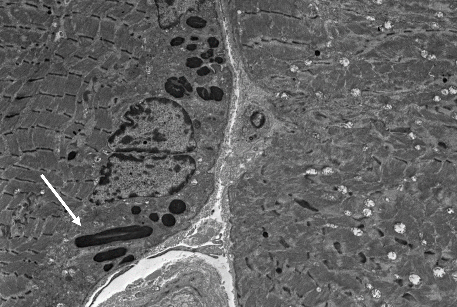
What are these dark (osmophilic) structures seen in this electron micrograph of skeletal muscle?
These osmophilic structures are Nemaline rods (largest, white arrow). These structures are classically seen in “Nemaline Myopathy” (NM; aka rod body myopathy). The rods are located in the sarcoplasm, and may also be seen within nuclei. NM is most commonly caused by dominant mutations of ACTA1 (encoding skeletal a-actin) and recessive mutations of NEB (encoding nebulin). Intranuclear rods are associated with pathogenic ACTA1 mutations. There are variable phenotypes of NM ranging from the severe congenital myopathy to adult-onset disease. Limited numbers of muscle fibers containing these structures may also be seen as an incidental finding in inflammatory myopathy (i.e. dermatomyositis), and near blood vessels or myotendinous insertion sites. Frequent nemaline rods may be seen in HIV myopathy or Sporadic Late Onset Nemaline Myopathy (SLONM) associated with monoclonal paraprotein. See references.
References:
Chahin N, Selcen D, Engel AG. Sporadic late onset nemaline myopathy. Neurology. 2005 Oct 25;65(8):1158-64. PMID: 16148261.
Colombo I, Scoto M, Manzur AY, et al. Congenital myopathies: natural history of a large pediatric cohort. Neurology. 2015 Jan 6;84(1):28-35. PMID: 25428687.
Dalakas MC, Pezeshkpour GH, Flaherty M. Progressive nemaline (rod) myopathy associated with HIV infection. N Engl J Med. 1987 Dec 17;317(25):1602-3. PMID: 3683496.
Sewry CA, Laitila JM, Wallgren-Pettersson C. Nemaline myopathies: a current view. J Muscle Res Cell Motil. 2019 Jun;40(2):111-126. PMID: 31228046.
Quick note: This post is to be used for informational purposes only and does not constitute medical or health advice. Each person should consult their own doctor with respect to matters referenced. Arkana Laboratories assumes no liability for actions taken in reliance upon the information contained herein.


