
This 6-week old baby presented with hypotonia at birth.
What are the structures indicated by arrows in Figures #1 and #2?
A. Angulated fibers
B. Macrophage
C. Motor end plates
D. Necrotic myofibers
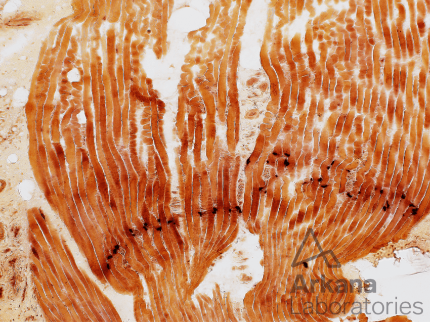
Figure 1: Esterase enzyme histochemical stain 100x original magnification
Low magnification image showing skeletal muscle in longitudinal orientation with bands of small dark staining areas (intense esterase activity).
Figure 2: Esterase enzyme histochemical stain 400x original magnification
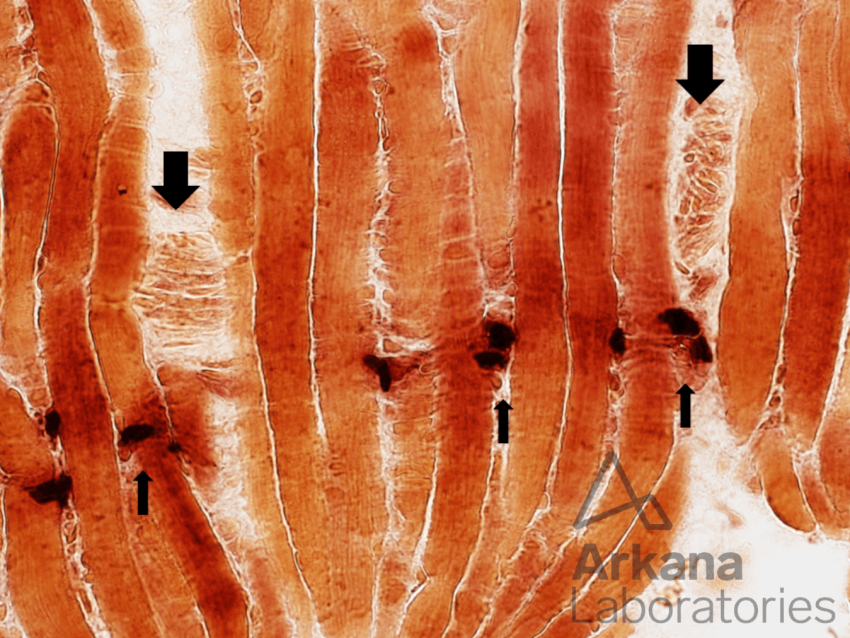
Higher magnification image showing small dark plaque-like staining areas (small arrows). Note also small intramuscular peripheral nerve twigs (larger arrows)
The correct answer is C. Motor End Plates
Neuromuscular junctions (NMJ) are most concentrated at the midbelly of muscles. This is nicely demonstrated in Figure #1 which highlights the band of NMJs in this longitudinally oriented frozen section of skeletal muscle.
Esterase enzyme histochemical stain demonstrates acetylcholinesterase activity concentrated within the neuromuscular junction.
Reference(s) / additional reading:
- Dubowitz V, Sewry C, Oldfors A. Muscle biopsy: A practical approach. St. Louis, MO: Elsevier Saunders; 2013.
- Blotnick-Rubin E, Anglister L. Fine Localization of Acetylcholinesterase in the Synaptic Cleft of the Vertebrate Neuromuscular Junction. Front Mol Neurosci. 2018 Apr 19;11:123. doi: 10.3389/fnmol.2018.00123. PMID: 29725289; PMCID: PMC5917012.
- https://www.ncbi.nlm.nih.gov/books/NBK470413/
- https://www.ncbi.nlm.nih.gov/pmc/articles/PMC5903580/
Quick note: This post is to be used for informational purposes only and does not constitute medical or health advice. Each person should consult their own doctor with respect to matters referenced. Arkana Laboratories assumes no liability for actions taken in reliance upon the information contained herein.