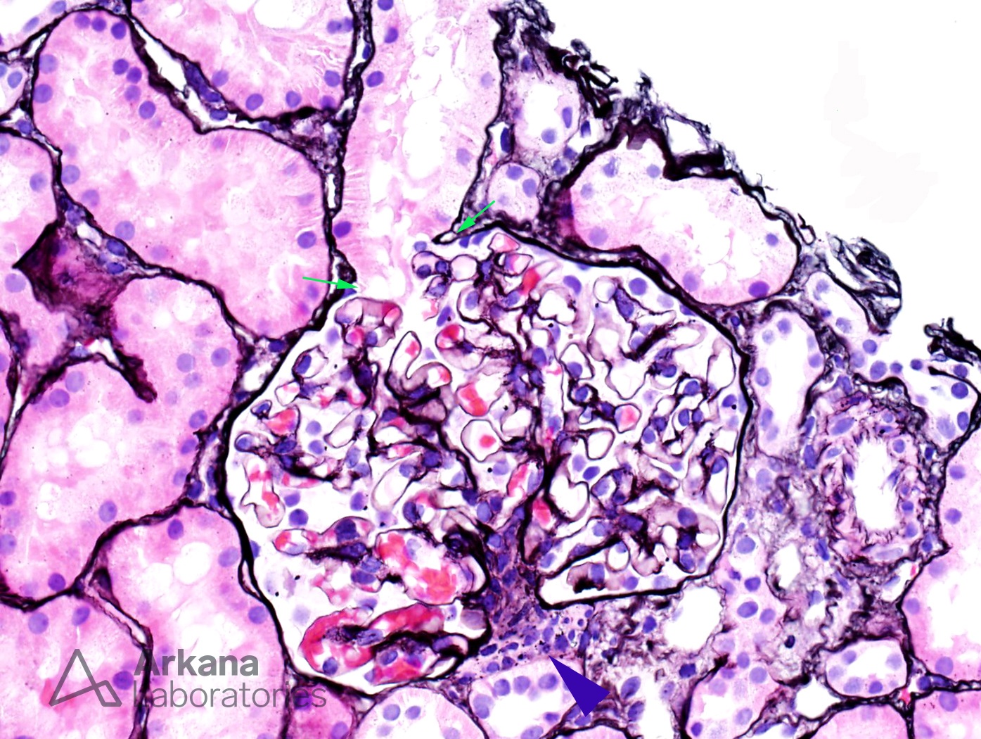
This is a normal glomerulus by light microscopy using the Jones silver stain. This section is fortuitously cut through the central plane of the spherical glomerulus. Note the proximal tubule arising at the top of the glomerulus, the so-called ‘tip area’ (green arrows). The hilum at the bottom of the photo contains the vascular entry and exit points as well as the juxtaglomerular apparatus (blue arrowhead). The glomerular basement membranes are delicate and without interruption. No endocapillary proliferation is present. The centrally located mesangial regions are not expanded and there is no mesangial hypercellularity. (Jones Silver 400x)
Quick note: This post is to be used for informational purposes only and does not constitute medical or health advice. Each person should consult their own doctor with respect to matters referenced. Arkana Laboratories assumes no liability for actions taken in reliance upon the information contained herein.