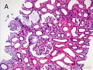
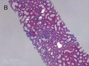
A 51-year-old female with a history of SLE was found to have acute renal failure. There was no evidence of glomerular proliferation by light microscopy but the biopsy did show (A) focal tubules with intraluminal and intracytoplasmic refractile crystals (arrows) (hematoxylin and eosin; original magnification × 100). (B) There are numerous intra-tubular birefringent crystals visible under polarized light (hematoxylin and eosin; original magnification × 50). These findings are consistent with kidney injury due to oxalate nephropathy. Known causes of oxalate nephropathy include primary hyperoxaluria, ethylene glycol intoxication, enteric hyperoxaluria (e.g. due to gastric bypass, chronic pancreatitis, small bowel resection, or malabsorption), exposure to the anesthetic agent methoxyflurane, vitamin B6 deficiency, and excessive ingestion of vitamin C or dietary substances rich in oxalic acid such as parsley, nuts, teas, and star fruit. The patient in the case shown here was found to be taking large daily doses of vitamin C.
Quick note: This post is to be used for informational purposes only and does not constitute medical or health advice. Each person should consult their own doctor with respect to matters referenced. Arkana Laboratories assumes no liability for actions taken in reliance upon the information contained herein.


