Clinical History
- This 35-year-old patient presented with headaches. MRI showed a sellar mass (diffuse enlargement of the pituitary gland) with expansion of the sella.
The figures are most consistent with which of the following?
A. Lymphocytic hypophysitis
B. Pituitary adenoma
C. Normal pituitary
D. Craniopharyngioma
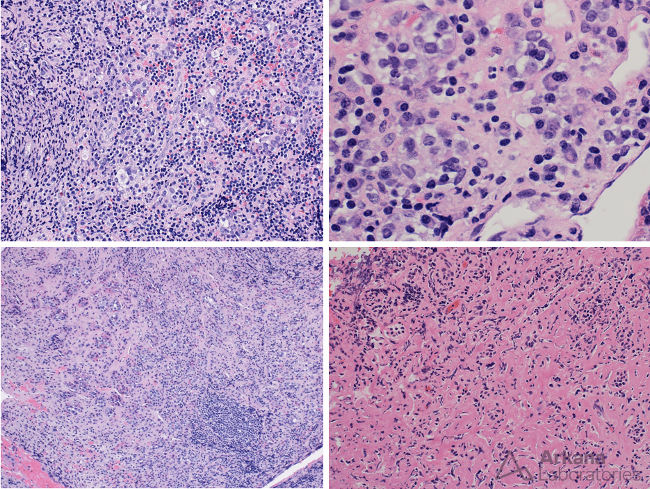
Figure 1:
- The images show variable chronic lymphoid inflammation with the patchy presence of mature appearing plasma cells (for example see arrows).
- Intact appearing nested adenohypophyseal cells are present (asterix).
- Focal fibrosis is noted.
- CD1a and OCT4, to evaluate for the possibility of Langerhans cell proliferation and paucicellular germinoma, were negative.
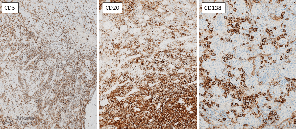
Figure 2:
- Focally greater than 10 IgG4 positive plasma cells were seen, but these were patchy in distribution and made up less than 20% of total IgG and/or CD138 positive plasma cells.
Correct Answer: A. Lymphocytic hypophysitis.
Reference(s) / additional reading:
- Naran J, Can AS. Lymphocytic Hypophysitis. [Updated 2023 Aug 8]. In: StatPearls [Internet]. Treasure Island (FL): StatPearls Publishing; 2023 Jan-. Available from: https://www.ncbi.nlm.nih.gov/books/NBK562255/
- Prete A, Salvatori R. Hypophysitis. 2021 Oct 15. In: Feingold KR, Anawalt B, Blackman MR, Boyce A, Chrousos G, Corpas E, de Herder WW, Dhatariya K, Dungan K, Hofland J, Kalra S, Kaltsas G, Kapoor N, Koch C, Kopp P, Korbonits M, Kovacs CS, Kuohung W, Laferrère B, Levy M, McGee EA, McLachlan R, New M, Purnell J, Sahay R, Shah AS, Singer F, Sperling MA, Stratakis CA, Trence DL, Wilson DP, editors. Endotext [Internet]. South Dartmouth (MA): MDText.com, Inc.; 2000–. PMID: 30160871.
Quick note: This post is to be used for informational purposes only and does not constitute medical or health advice. Each person should consult their own doctor with respect to matters referenced. Arkana Laboratories assumes no liability for actions taken in reliance upon the information contained herein.


