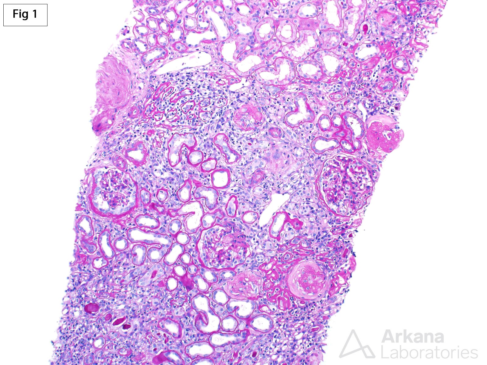
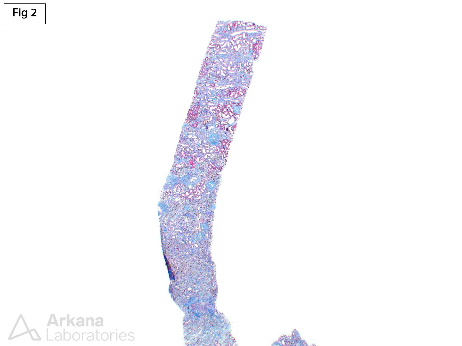
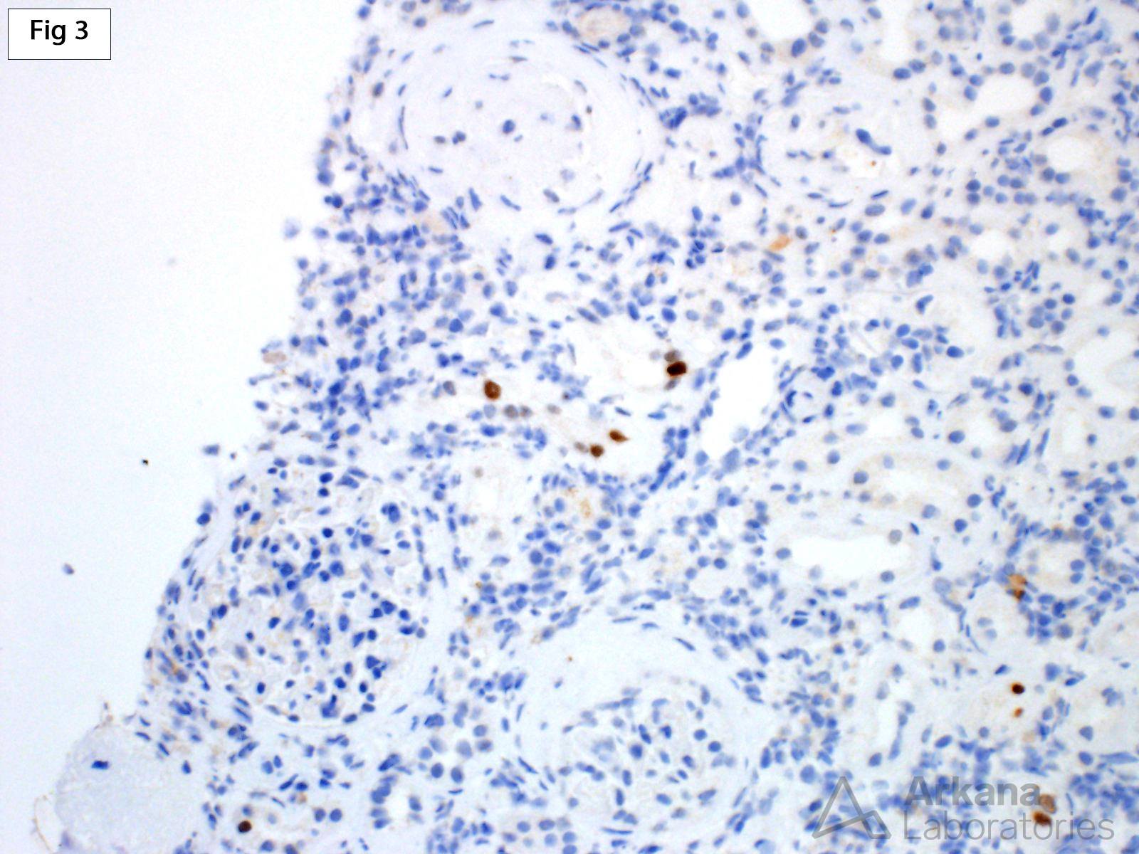
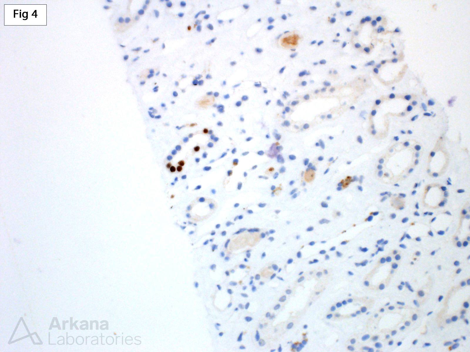
Polyomavirus nephropathy (PVN), predominantly caused by BK virus reactivation in the urogenital tract, is an important complication of renal allografts (reference 1). While it rarely affects the native kidney, it may be seen in the setting of chronic immunosuppression, including bone marrow transplantation, HIV infection, chemotherapy and in the setting of other solid organ transplants (reference 2). The case shown is from a 69-year-old female with a history of sarcoidosis and cirrhosis who is now 5 years post liver transplant. The patient is currently being evaluated for a progressive increase in serum creatinine. The biopsy shows significant, non-specific tubulointerstitial scarring with mild mixed inflammation (Fig 1 and 2). The tubular epithelium in the affected areas shows mild nuclear reactive changes, but no definitive viral inclusions are identified. An SV40 immunoperoxidase stain shows positive nuclear staining involving the cortex and medulla (Fig 3 and 4), diagnostic of PVN.
References:
1. Nickeleit V, Singh HK, Randhawa P, et al. The Banff Working Group Classification of Definitive Polyomavirus Nephropathy: Morphologic Definitions and Clinical Correlations. J Am Soc Nephrol. 2018;29:680-693.
2. Sharma SG, Nickeleit V, Herlitz LC, et al. BK polyomavirus nephropathy in the native kidney. Nephrol Dial Transplant. 2013;28:620-31.
Quick note: This post is to be used for informational purposes only and does not constitute medical or health advice. Each person should consult their own doctor with respect to matters referenced. Arkana Laboratories assumes no liability for actions taken in reliance upon the information contained herein.


