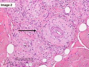
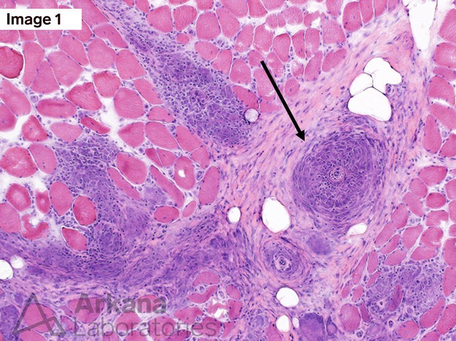
What is your diagnosis and how would you characterize the designated lesions in Figures 1 & 2? (Black Arrows)

Hematoxylin and eosin stained FFPE tissue section (200x original magnification
Granulomatous myopathy, most consistent with Sarcoidosis (so-called “Sarcoid Myopathy”). The granulomatous inflammation of Sarcoidosis may show a perivascular distribution and may cause vascular compromise (vasculopathy or vasculitis). Of note, Sarcoidosis is a diagnosis of exclusion (i.e. other causes of granulomatous inflammation must be excluded). See references.
References:
Cohen Aubart F, Abbara S, Maisonobe T, et al. Symptomatic muscular sarcoidosis: Lessons from a nationwide multicenter study. Neurol Neuroimmunol Neuroinflamm. 2018 Mar 16;5(3):e452. PMID: 29845092.
Le Roux K, Streichenberger N, Vial C, et al. Granulomatous myositis: a clinical study of thirteen cases. Muscle Nerve. 2007 Feb;35(2):171-7. PMID: 17068767.
Ungprasert P, Ryu JH, Matteson EL. Clinical Manifestations, Diagnosis, and Treatment of Sarcoidosis. Mayo Clin Proc Innov Qual Outcomes. 2019 Aug 2;3(3):358-375. PMID: 31485575.
Quick note: This post is to be used for informational purposes only and does not constitute medical or health advice. Each person should consult their own doctor with respect to matters referenced. Arkana Laboratories assumes no liability for actions taken in reliance upon the information contained herein.


