What pathologic process is demonstrated by the low and higher magnification images of a teased nerve preparation?
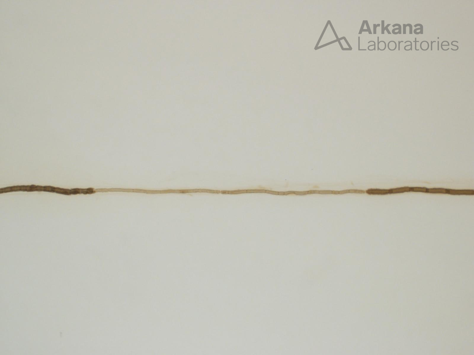
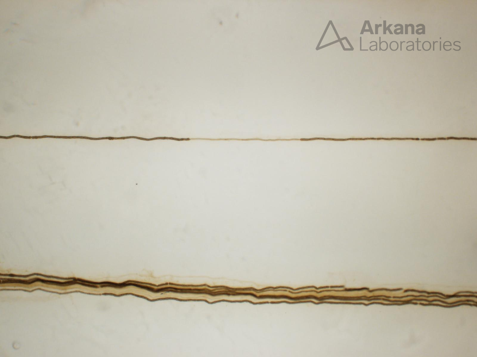
The image shows a large diameter myelinated axon with focal short and thinly myelinated internodes. This alteration indicates prior segmental demyelination and subsequent successful remyelination.
Quick note: This post is to be used for informational purposes only and does not constitute medical or health advice. Each person should consult their own doctor with respect to matters referenced. Arkana Laboratories assumes no liability for actions taken in reliance upon the information contained herein.