Clinical History
- This 50-year-old patient presented with muscle weakness and stiffness. They had recently been identified to have diffuse thickening of the stomach wall.
Based on Figures #1 – #4 which of the following is the most likely diagnosis?
A. Macrophagic myofasciitis
B. Lymphoma
C. Signet ring carcinoma
D. Inflammatory myopathy
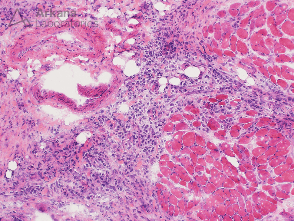
Figure 1: Medium magnification image shows abnormal cellularity within the perimysium with infiltrates into the periphery of some muscle fascicles. At this magnification, the cells seem somewhat more atypical than what would be typically seen in chronic lymphoid inflammation.
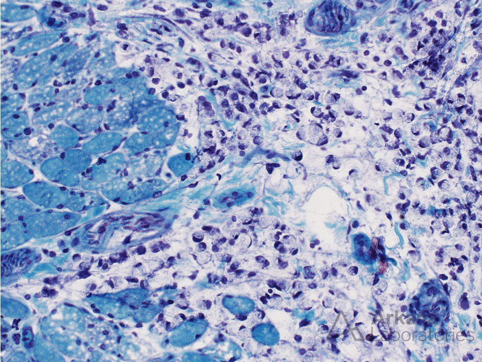
Figure 2: Higher magnification image shows that the atypical cells have moderate amounts of vacuolated cytoplasm that displaces the nucleus to the periphery (“signet ring” appearance). Note that the nuclei of the atypical cells are larger than the myofiber nuclei.
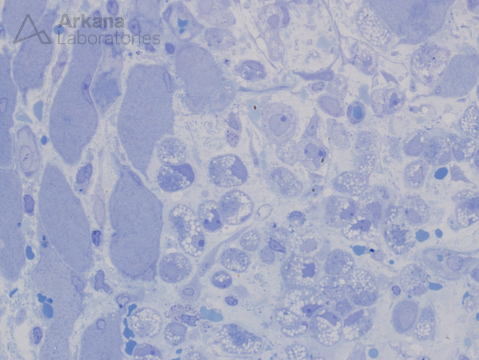
Figure 3: High magnification image showing that the atypical cells (see arrows) have moderate amounts of vacuolated cytoplasm that displaces the nucleus to the periphery (“signet ring” appearance). The nuclei are moderately enlarged and have readily visible nucleoli..
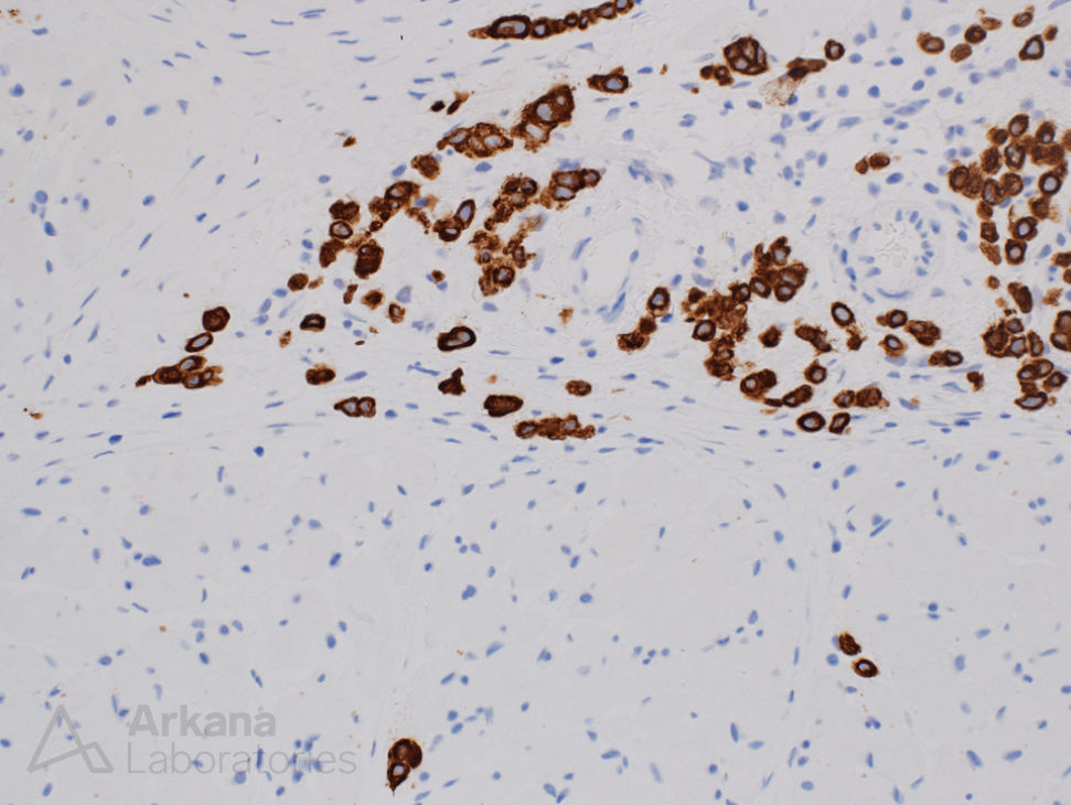
Figure 4: Higher magnification image showing that the atypical cells show strong diffuse cytoplasmic staining for cytokeratin.
Correct Answer: C
- The correct answer is signet ring carcinoma. In the context of this patient’s clinical history, a gastric primary is suspected. However, additional studies (immunohistochemical stains) would be required to rule out other primary sites (for example lobular carcinoma of breast).
Quick note: This post is to be used for informational purposes only and does not constitute medical or health advice. Each person should consult their own doctor with respect to matters referenced. Arkana Laboratories assumes no liability for actions taken in reliance upon the information contained herein.


