Clinical History
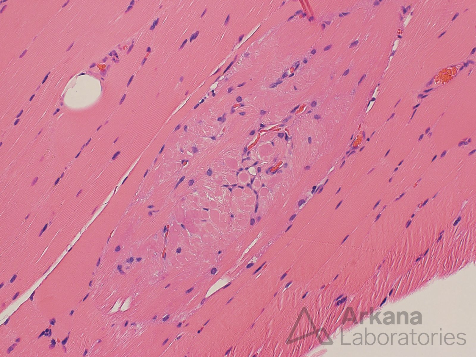
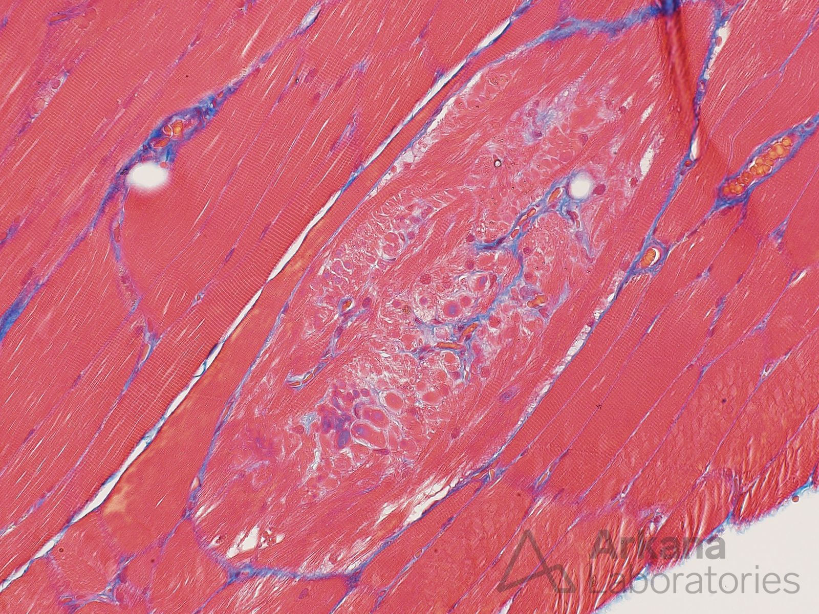
This 45-year-old patient underwent muscle biopsy to evaluate for muscle weakness. The biopsy showed denervation type changes (neurogenic atrophy). An unusual looking muscle fiber was noted on paraffin sections.
Based on the H&E and Masson Trichrome stained section images what relatively common morphologic abnormality is this most likely to represent?
Answer:
- This is felt to most likely represent a hypertrophic muscle fiber with so-called muscle fiber splitting seen in longitudinal orientation. Note the fine capillaries highlighted in blue by the Masson Trichrome stain.
- Recent evidence suggests that myofiber splitting or branching may be due to myotube fusion following myofiber necrosis.
Reference(s)/Additional Reading:
- Højfeldt G, Sorenson T, Gonzales A, Kjaer M, Andersen JL, Mackey AL. Fusion of myofibre branches is a physiological feature of healthy human skeletal muscle regeneration. Skelet Muscle. 2023 Aug 12;13(1):13. doi: 10.1186/s13395-023-00322-2. PMID: 37573332; PMCID: PMC10422711.
- Swash M, Schwartz MS. Implications of longitudinal muscle fibre splitting in neurogenic and myopathic disorders. J Neurol Neurosurg Psychiatry. 1977 Dec;40(12):1152-9. doi: 10.1136/jnnp.40.12.1152. PMID: 591984; PMCID: PMC492938.
- Murach KA, Dungan CM, Peterson CA, McCarthy JJ. Muscle Fiber Splitting Is a Physiological Response to Extreme Loading in Animals. Exerc Sport Sci Rev. 2019 Apr;47(2):108-115. doi: 10.1249/JES.0000000000000181. PMID: 30640746; PMCID: PMC6422761.
Quick note: This post is to be used for informational purposes only and does not constitute medical or health advice. Each person should consult their own doctor with respect to matters referenced. Arkana Laboratories assumes no liability for actions taken in reliance upon the information contained herein.