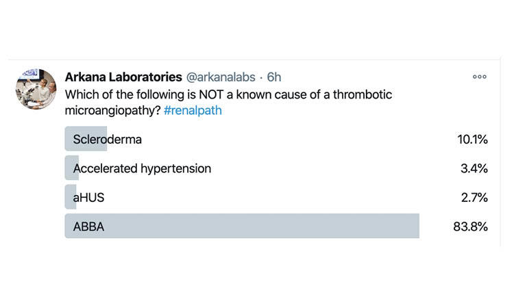
Answer: D
The histologic features of a thrombotic microangiopathy (TMA) are non-specific as to etiology and include both acute and chronic changes. Acute changes of TMA include glomeruli demonstrating mesangiolysis, endothelial swelling, as well as intracapillary fibrin thrombi, and fragmented red blood cells. Blood vessels in the acute phase will also classically show endothelial swelling along with intimal mucoid edema and entrapped schistocytes. Chronic TMA changes include glomerular basement duplication with double contour formation and arterial/arteriolar intimal fibrosis with concentric lamination (onion skinning), to name a few. All of the above are known to lead to TMA except for anti-brush border antibody disease (ABBA).
Quick note: This post is to be used for informational purposes only and does not constitute medical or health advice. Each person should consult their own doctor with respect to matters referenced. Arkana Laboratories assumes no liability for actions taken in reliance upon the information contained herein.

